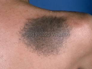
Nevus of Ito is a unilateral, benign, dermal melanocytosis that presents as a darkly pigmented patch on the side of the neck and shoulder in the distribution of the posterior supraclavicular and lateral cutaneous brachial nerves.
It shares clinical and histopathologic features, aside from location, with other dermal melanocytoses including nevus of Ota (trigeminal distribution), congenital dermal melanocytosis (formerly known as a Mongolian spot in the lumbosacral distribution), and dermal melanocyte hamartoma. Dermal melanocytoses are caused by an increased number of dermal dendritic melanocytes and can be congenital or acquired. All dermal melanocytoses are more often seen in patients of Asian or African descent, and nevus of Ito, in particular, is more common among females.
Unlike the more common congenital dermal melanocytosis, nevus of Ito lesions do not spontaneously regress and may darken or grow in size with puberty. These changes are thought to be secondary to ultraviolet radiation and hormonal influences. Patients have noted sensory changes in the area of the nevus of Ito. Extremely rarely, melanoma arising in a nevus of Ito has been reported.
Nevus of Ito in Infant/Neonate
Synopsis

Codes
ICD10CM:
D22.9 – Melanocytic nevi, unspecified
SNOMEDCT:
48543002 – Nevus of Ito
D22.9 – Melanocytic nevi, unspecified
SNOMEDCT:
48543002 – Nevus of Ito
Look For
Subscription Required
Diagnostic Pearls
Subscription Required
Differential Diagnosis & Pitfalls

To perform a comparison, select diagnoses from the classic differential
Subscription Required
Best Tests
Subscription Required
Management Pearls
Subscription Required
Therapy
Subscription Required
References
Subscription Required
Last Updated:06/07/2016
 Patient Information for Nevus of Ito in Infant/Neonate
Premium Feature
VisualDx Patient Handouts
Available in the Elite package
- Improve treatment compliance
- Reduce after-hours questions
- Increase patient engagement and satisfaction
- Written in clear, easy-to-understand language. No confusing jargon.
- Available in English and Spanish
- Print out or email directly to your patient
Upgrade Today

Nevus of Ito in Infant/Neonate