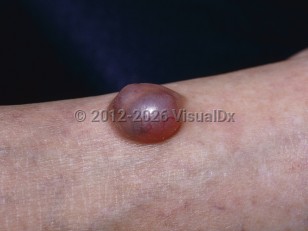
Nodular hidradenoma is a rare, benign adnexal tumor that displays apocrine differentiation in the majority of cases (clear cell hidradenoma) and eccrine differentiation (poroid hidradenoma) in the remainder. The two histopathologic types are indistinguishable clinically. A 0.5-3 cm firm nodule develops on the head, anterior trunk, or upper limbs, or any other cutaneous site less frequently. It grows slowly and may ulcerate.
Nodular hidradenoma is more common in females and can occur in any ethnicity and at any age including infancy, although it is most common in the fourth decade of life. There are currently no known risk factors. Local recurrence can also occur, especially with inadequate surgical excision.
Hidradenocarcinoma (also known as malignant nodular hidradenoma [MNH]) is a very rare malignant tumor that can arise from nodular hidradenoma, although it typically arises de novo.
Nodular hidradenoma
Alerts and Notices
Important News & Links
Synopsis

Codes
ICD10CM:
D23.9 – Other benign neoplasm of skin, unspecified
SNOMEDCT:
253020008 – Hidradenoma of skin
D23.9 – Other benign neoplasm of skin, unspecified
SNOMEDCT:
253020008 – Hidradenoma of skin
Look For
Subscription Required
Diagnostic Pearls
Subscription Required
Differential Diagnosis & Pitfalls

To perform a comparison, select diagnoses from the classic differential
Subscription Required
Best Tests
Subscription Required
Management Pearls
Subscription Required
Therapy
Subscription Required
References
Subscription Required
Last Updated:06/01/2023
Nodular hidradenoma