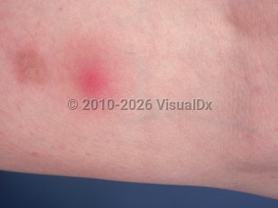
Nodular vasculitis is a form of lobular panniculitis with multiple etiologies. It presents with tender erythematous or violaceous nodules and/or plaques, usually on the calves. Ulceration and drainage may occur. Lesions tend to heal with scarring and are prone to recurrence. Women are most commonly affected (80%-90% of patients), with peaks in incidence during early adolescence and around menopause.
There are many etiologies. Idiopathic nodular vasculitis may be seen on the calves of women in middle age who have varicosities. Mycobacterium tuberculosis infection is one of the strongest associated triggers. The term erythema induratum (of Bazin) refers specifically to cases of nodular vasculitis associated with underlying tuberculosis. Cases can also be induced by infectious agents such as Nocardia, Pseudomonas, Chlamydia, or hepatitis B or hepatitis C infections. Drugs may also be causative agents, with tumor necrosis factor (TNF)-alpha inhibitors, notably etanercept, implicated in case reports. The condition is thought to be either an immune complex-mediated vasculitis or a type IV cell-mediated response to an antigenic stimulus.
Related topic: cutaneous tuberculosis
Nodular vasculitis
Alerts and Notices
Important News & Links
Synopsis

Codes
ICD10CM:
L95.8 – Other vasculitis limited to the skin
SNOMEDCT:
55275006 – Nodular vasculitis
L95.8 – Other vasculitis limited to the skin
SNOMEDCT:
55275006 – Nodular vasculitis
Look For
Subscription Required
Diagnostic Pearls
Subscription Required
Differential Diagnosis & Pitfalls

To perform a comparison, select diagnoses from the classic differential
Subscription Required
Best Tests
Subscription Required
Management Pearls
Subscription Required
Therapy
Subscription Required
Drug Reaction Data
Subscription Required
References
Subscription Required
Last Reviewed:08/15/2022
Last Updated:08/29/2022
Last Updated:08/29/2022
Nodular vasculitis