Ocular pterygium - External and Internal Eye
Alerts and Notices
Important News & Links
Synopsis

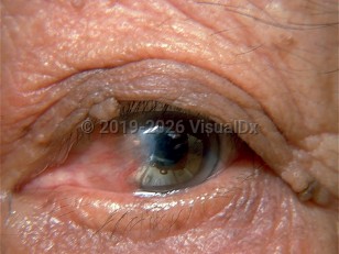
A pterygium is a soft, fleshy benign growth on the bulbar surface of the eye in the perilimbal area, which may extend onto the corneal surface. These growths can vary from nearly flat and atrophic to large, rapidly growing fibrovascular lesions that can distort the corneal topography and even reach the optical center of the cornea. Most often they are found in the nasal interpalpebral part of the bulbar conjunctiva but can occur temporally as well. They are frequently bilateral yet unequal in size. The incidence is higher in men than in women and is far more frequent in warm or tropical climates. Patients present with either just the finding of the growth or complain of irritation, redness, blurring of vision, itching, difficulty wearing contact lenses, and/or foreign body feeling. Exposure to UV light, allergens, noxious chemicals, and irritants such as wind, dirt, dust, and air pollution all contribute to the etiology of pterygiums. There may also be a genetic component as well.
Codes
ICD10CM:
H11.009 – Unspecified pterygium of unspecified eye
SNOMEDCT:
77489003 – Pterygium
H11.009 – Unspecified pterygium of unspecified eye
SNOMEDCT:
77489003 – Pterygium
Look For
Subscription Required
Diagnostic Pearls
Subscription Required
Differential Diagnosis & Pitfalls

To perform a comparison, select diagnoses from the classic differential
Subscription Required
Best Tests
Subscription Required
Management Pearls
Subscription Required
Therapy
Subscription Required
References
Subscription Required
Last Updated:04/30/2017
 Patient Information for Ocular pterygium - External and Internal Eye
Premium Feature
VisualDx Patient Handouts
Available in the Elite package
- Improve treatment compliance
- Reduce after-hours questions
- Increase patient engagement and satisfaction
- Written in clear, easy-to-understand language. No confusing jargon.
- Available in English and Spanish
- Print out or email directly to your patient
Upgrade Today

Ocular pterygium - External and Internal Eye