Contents

SynopsisCodesLook ForDiagnostic PearlsDifferential Diagnosis & PitfallsBest TestsManagement PearlsTherapyReferences

Information for Patients

View all Images (23)

Oral hemangioma - Oral Mucosal Lesion
Other Resources UpToDate PubMed

Oral hemangioma - Oral Mucosal Lesion

Contributors: Carl Allen DDS, MSD, Sook-Bin Woo MS, DMD, MMSc
Other Resources UpToDate PubMed

Synopsis

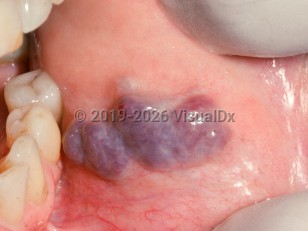
True oral hemangioma is a developmental vascular malformation of capillaries. They tend to affect infants and young children with a slight female predilection. The lesions are not painful but will bleed if traumatized. They are usually present, but subtle, at birth, grow rapidly within the first few years of life, and then involute.

The more common vascular malformations in the oral cavity, loosely called hemangiomas, are proliferations of capillaries or venules that do not involute but persist and grow very slowly over years. They tend to occur on the tongue, buccal mucosa, and labial mucosa. The intrabony lesions are best classified as "vascular malformations."

Codes

ICD10CM:
D10.30 – Benign neoplasm of unspecified part of mouth

SNOMEDCT:
403963001 – Hemangioma of oral cavity

Look For

Subscription Required

Diagnostic Pearls

Subscription Required

Differential Diagnosis & Pitfalls

  • Lobular capillary hemangioma (pyogenic granuloma) is a reactive proliferation of blood vessels that occurs in adults and during pregnancy. It is usually present on the gingiva, buccal mucosa, or labial mucosa.
  • Peripheral giant cell granuloma
  • Kaposi sarcoma may present as a purplish-blue nodule with a tendency to bleed. It is seen in the oral cavity, primarily in those with AIDS.
  • Oral varices
  • Some abscesses may appear purplish-red, but these are always associated with an infectious process, odontogenic or otherwise.
  • Leukemic infiltration
  • Large varices may appear similar but generally occur in older individuals and are common on the lip.
  • Sturge-Weber syndrome is a vascular malformation involving the soft tissues of the face and oral mucosa that follows the distribution of the trigeminal nerve. If the ophthalmic division is affected, there is usually concomitant meningeal involvement. Oral lesions are diffuse, purple plaques, and the overlying skin has a port-wine stain.
  • Granulomatosis with polyangiitis
  • Squamous cell carcinoma

Best Tests

Subscription Required

Management Pearls

Subscription Required

Therapy

Subscription Required

References

Subscription Required

Last Updated:10/30/2017
Copyright © 2023 VisualDx®. All rights reserved.
Patient Information for Oral hemangioma - Oral Mucosal Lesion
Print E-Mail Images (23)
Contributors: Medical staff writer

Overview

A hemangioma is an area of soft tissue that appears purple or dark red due to small blood vessels or capillaries that are malformed. Oral hemangiomas can appear on the lips, tongue, and mucous membrane of the mouth. They are usually painless, but they can bleed when bumped or irritated. They may cause swelling if they develop in the jawbone.

Who’s At Risk

Oral hemangiomas are common in infants and children, but they can occur in adults as well. They may grow rapidly in infants, and are harmless (benign) and painless, unless irritated. Adults may develop oral hemangiomas over a period of years.

Signs & Symptoms

Oral hemangiomas look like red, blue, or purple patches on the skin or other soft tissue, particularly in the mouth or on the tongue or lips. They are painless, but they can bleed if irritated or traumatized.

Self-Care Guidelines

Avoid poking or injuring the oral hemangioma, as it may bleed.

Most oral hemangiomas will resolve on their own, without treatment.

When to Seek Medical Care

If your child has an oral hemangioma that interferes with talking or eating, see your pediatrician.

Treatments

Oral hemangiomas do not need treatment, unless bleeding has become a problem. Hemangiomas that interfere with breathing, swallowing, eating, etc. may require removal. Hemangiomas can also be removed for cosmetic reasons. Many hemangiomas will go away on their own.
Copyright © 2023 VisualDx®. All rights reserved.
Oral hemangioma - Oral Mucosal Lesion
A medical illustration showing key findings of Oral hemangioma : Oral vascular mass, Purple color
Clinical image of Oral hemangioma - imageId=2505948. Click to open in gallery.  caption: 'Confluent violaceous nodules on the buccal mucosa.'
Confluent violaceous nodules on the buccal mucosa.
Copyright © 2023 VisualDx®. All rights reserved.