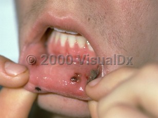
Oral leukemic infiltration is the presence of localized or generalized tissue hyperplasia of the mouth caused by collections of malignant white blood cells or their precursors. The demographic features follow that of the particular leukemia. However, the most common presentation is leukemic infiltration of the gingiva in acute monocytic or acute myelomonocytic leukemia, a disease of adults. Lesions are generally not painful, although they may bleed. Because of immunosuppression secondary to the leukemia, lesions may also exhibit secondary infection by viral or fungal agents.
Patients report weight loss, fatigue, loss of appetite, and easy bruisability. The infiltrate often develops over weeks and months.
Oral leukemic infiltration - Oral Mucosal Lesion
Alerts and Notices
Important News & Links
Synopsis

Codes
ICD10CM:
C95.90 – Leukemia, unspecified not having achieved remission
SNOMEDCT:
404156009 – Leukemic infiltration of skin
C95.90 – Leukemia, unspecified not having achieved remission
SNOMEDCT:
404156009 – Leukemic infiltration of skin
Look For
Subscription Required
Diagnostic Pearls
Subscription Required
Differential Diagnosis & Pitfalls

To perform a comparison, select diagnoses from the classic differential
Subscription Required
Best Tests
Subscription Required
Management Pearls
Subscription Required
Therapy
Subscription Required
References
Subscription Required
Last Updated:01/24/2008
Oral leukemic infiltration - Oral Mucosal Lesion