Oral melanoacanthoma (OM) is a benign acquired pigmentation of the oral mucosa that is seen almost exclusively in Black individuals, with a female predilection, and is most common in the third and fourth decades of life. While the pathogenesis of this lesion is not fully understood, OM is thought to be reactive in nature, arising in response to chronic trauma or chemical irritants.
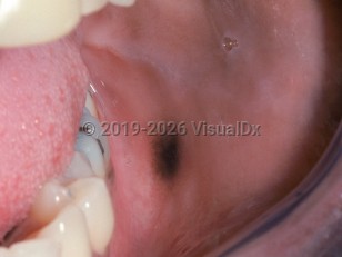
OM presents as a well-demarcated, smooth macule or slightly elevated plaque with a relatively uniform dark brown to black color. Its size typically ranges from 0.5 to 3 cm in diameter. OM is most often solitary, but multifocal lesions have been reported. The buccal mucosa is the most common site of involvement. Occasionally, patients may develop a synchronous melanoacanthoma on the contralateral buccal mucosa. OM has also been reported on the lip, palate, and gingiva. Multifocal OM is most commonly seen on the palate. While OM is often asymptomatic, pruritus and/or pain may be present.
Patients who have been aware of the development of this lesion often describe a relatively rapid increase in size over a period of a few weeks. Spontaneous resolution has been reported if the inciting factor is withdrawn or after biopsy.
Oral melanoacanthoma - Oral Mucosal Lesion
Alerts and Notices
Important News & Links
Synopsis

Codes
ICD10CM:
D10.30 – Benign neoplasm of unspecified part of mouth
SNOMEDCT:
394727000 – Melanoacanthoma
D10.30 – Benign neoplasm of unspecified part of mouth
SNOMEDCT:
394727000 – Melanoacanthoma
Look For
Subscription Required
Diagnostic Pearls
Subscription Required
Differential Diagnosis & Pitfalls

To perform a comparison, select diagnoses from the classic differential
Subscription Required
Best Tests
Subscription Required
Management Pearls
Subscription Required
Therapy
Subscription Required
References
Subscription Required
Last Reviewed:10/07/2021
Last Updated:10/07/2021
Last Updated:10/07/2021
Oral melanoacanthoma - Oral Mucosal Lesion