Pancreatic panniculitis is also known as subcutaneous fat necrosis, enzymatic panniculitis, or pancreatic fat necrosis. It is an uncommon cutaneous complication that occurs in 2%-3% of patients with pancreatic disease.
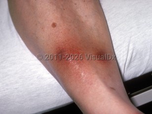
The condition presents as tender, red, erythematous subcutaneous nodules, most frequently on the lower extremities, although they may occur on the breasts, upper extremities, trunk, and buttocks. Long-standing subcutaneous nodules often ulcerate spontaneously and drain a yellow to brown oily material. Given the propensity for this panniculitis to ulcerate, cases have often been misdiagnosed as an infectious process such as necrotizing fasciitis and have thus undergone unnecessary surgical debridement. The distal lower extremities, notably around the ankles and knees, are the most common locations for nodules to ulcerate. Necrosis of the periarticular fat may rarely occur and, in this setting, patients may also experience arthritis involving the ankles and knees. Even rarer associations include necrosis of abdominal fat or bone marrow fat, pleural effusions, mesenteric thrombosis, and eosinophilia.
Pancreatic panniculitis occurs in the setting of acute or chronic pancreatitis and in the context of pancreatic acinar carcinoma, where panniculitis may herald metastatic disease. Associations with other types of pancreatic cancer, such as islet cell carcinoma, have been reported but occur less frequently. Other less common associations include pancreas divisum, pancreatic pseudocysts, vasculopancreatic fistulas, traumatic pancreatitis, and pancreatitis secondary to sulindac intake. Cases have been reported following endoscopic retrograde cholangiopancreatography (ERCP). Pancreatic panniculitis has also been reported with nonpancreatic entities such as intrahepatic cholangiocarcinoma.
Pancreatic panniculitis can be a presenting feature of subclinical pancreatitis in which elevated amylase and lipase may be the only laboratory abnormalities. It is thought that release of trypsin, lipase, amylase, and other lipolytic enzymes from the pancreas into the circulation results in formation of triglycerides from glycerol esters in the pannus, which leads to necrosis.
Cutaneous disease can precede or follow underlying pancreatic disease; sometimes the cutaneous eruption precedes the typical presentation of pancreatitis by weeks to months.
Resolution of the nodules with scarring may occur within weeks to months after treatment of pancreatitis or after resection of pancreatic carcinoma.
Immunocompromised Patient Considerations:
A single case was reported in a patient with primary HIV infection and a hemophagocytic syndrome.
Pediatric Patient Considerations:
There is one case report of a child with systemic lupus erythematosus treated with prednisone, azathioprine, and hydrochlorothiazide who developed pancreatitis and associated subcutaneous nodular fat necrosis and calcinosis cutis.
Pancreatic panniculitis
Alerts and Notices
Important News & Links
Synopsis

Codes
ICD10CM:
M79.3 – Panniculitis, unspecified
SNOMEDCT:
403416005 – Panniculitis secondary to pancreatic disease
M79.3 – Panniculitis, unspecified
SNOMEDCT:
403416005 – Panniculitis secondary to pancreatic disease
Look For
Subscription Required
Diagnostic Pearls
Subscription Required
Differential Diagnosis & Pitfalls

To perform a comparison, select diagnoses from the classic differential
Subscription Required
Best Tests
Subscription Required
Management Pearls
Subscription Required
Therapy
Subscription Required
Drug Reaction Data
Subscription Required
References
Subscription Required
Last Reviewed:10/14/2020
Last Updated:10/25/2020
Last Updated:10/25/2020
Pancreatic panniculitis