At the immediate point of care:
- CPR if needed.
- Stabilize the neck if a neck injury is suspected.
- Stabilize the child and provide supportive care.
- Assess the patient for additional life-threatening injuries (intra-abdominal and intrathoracic), as you would any trauma patient.
- Monitor for signs of worsening of intracranial hemorrhage, herniation, and neurologic deterioration.
- Ventilatory support may be required.
Abusive head trauma, also known as shaken baby syndrome, shaken impact syndrome, inflicted head injury, and whiplash shaken infant syndrome, is a form of trauma that arises when a baby (or small child) is shaken and/or struck so violently that it leads to cerebral injury.
Inconsolable crying is usually the trigger for such actions on the part of the caregiver. Victims are typically between 2 and 8 months of age, although this type of injury has been seen in older children. All races and ethnicities as well as levels of socioeconomic status are affected, but physicians may be less likely to recognize abusive head injury when the caregivers are White and from a higher socioeconomic class, and when the child comes from an intact family.
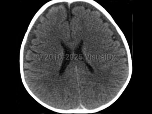
It is thought that the primary injury arises as a result of the multidirectional forces from shaking, leading to a form of whiplash injury associated with diffuse axonal injury, tearing of bridging veins, and subsequent intracranial bleeding. The secondary brain injury is due to a combination of hypoxia, ischemia, and brain edema. The end result is destruction of brain cells, a reduced supply of oxygen to brain cells, and brain swelling leading to varying degrees of neurological deterioration and occasionally death. Anatomically, the combination of a not-yet myelinated brain, a relatively heavy head upon weak neck muscles, and lack of control of the head and neck are factors that predispose babies to this form of injury.
Often, no history of trauma is offered, or there is a history of a short fall or a history that a sibling injured the infant. The degree of injury is not compatible with the history given.
Abusive head trauma may present with one or more of the following clinical features: subdural hemorrhage, retinal hemorrhage, and encephalopathy. Retinal hemorrhages are present in approximately 85% of cases and may be unilateral, but the diagnosis of abusive head trauma should not be excluded if they are absent.
The degree of damage inflicted depends on the force of shaking and the length of time the baby was shaken. On presentation, the baby may appear lethargic, irritable, or in a comatose state; have a bulging anterior fontanel; be bradycardic and hypothermic; and seem to have difficulty breathing.
Symptoms of abusive head trauma may be nonspecific and include:
- Irritability.
- A brief resolved unexplained event (BRUE).
- Seizures.
- Difficulty breathing / apnea, which may be severe enough to require intubation.
- Poor feeding.
- Lethargy.
- Loss of consciousness / comatose state.
- Vomiting.
- Rigidity / hypertonia.
- Inability to lift the head / hypotonia.
- Size of head appears larger than usual if there have been prior episodes.
Following episodes of infant shaking, the consequences can vary from no detectable adverse effects to death. The fatality rate following this type of trauma is high, at approximately 20%, and the prognosis for those who survive is variable. Even when infants survive, there remains the possibility of lifelong disabilities that include cerebral palsy, intellectual disability, blindness, paralysis, epilepsy, speech delay, hearing impairment, and learning disabilities.
Injuries that raise concern for abusive head trauma – such as severe intracranial injury, with or without retinal hemorrhages, with or without additional injuries, and having an inadequate trauma history to explain the findings – have not been documented in falls from short heights or from routine handling or play with infants such as throwing a baby into the air and catching them.
Related topic: child physical abuse