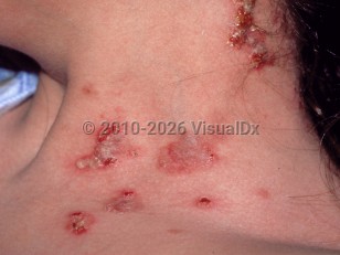
Pemphigus vegetans (PVeg), a rare variant of pemphigus vulgaris (PV), is an autoimmune bullous disease characterized by vegetating lesions in intertriginous areas. As in PV, circulating immunoglobulin G (IgG) autoantibodies against desmoglein, a keratinocyte cell surface molecule, lead to disruption of normal cell adhesion between keratinocytes and subsequent formation of vesicles and bullae. Desmocollins have been noted to be additional autoantibody targets in PVeg.
PVeg is rare and makes up an estimated 2%-5% of the total cases of pemphigus. Although this disease can affect people of all age groups, it occurs primarily during middle age with a median onset of 40-60 years. It affects males and females equally.
PVeg has been divided into two subtypes depending on clinical presentation and disease course:
The Neumann type is characterized by flaccid bullae and erosions in intertriginous areas that develop overlying vegetations. This subtype is more common, with a clinical course that parallels PV.
The Hallopeau type has a more benign course and is characterized by erupting crops of pustules that evolve into crusted plaques. It may remit spontaneously.
Mucous membrane involvement is a frequent occurrence. The scalp may also manifest vegetative plaques. The perioral area is a site of predilection in the Hallopeau type.
Codes
ICD10CM: L10.1 – Pemphigus vegetans
SNOMEDCT: 81285006 – Pemphigus vegetans
Look For
Subscription Required
Diagnostic Pearls
Subscription Required
Differential Diagnosis & Pitfalls
Generalized PV – Similar in presentation to Neumann type but not limited to intertriginous areas and without vegetating plaques.
Paraneoplastic pemphigus – Associated with malignancy, most commonly non-Hodgkin lymphoma; mucous membranes are the site of most severe involvement.
IgA pemphigus – Mucous membranes are spared; immunoglobulin A (IgA) autoantibodies versus IgG in PVeg.
Drug-induced pemphigus – Most commonly captopril and D-penicillamine implicated; a precise drug history is critical, as these can be difficult to distinguish, especially if only oral lesions are present. Pemphigus foliaceus is more common than PV in cases of drug-induced pemphigus.
Pemphigus foliaceus (PF) – Mucous membranes are spared; scales and crusts over erythematous skin in seborrheic areas.
Pemphigus erythematosus (also known as Senear-Usher syndrome) is a localized variety of PF largely confined to the malar region and seborrheic sites.
Fogo selvagem is an endemic form of PF found in rural Brazil.
Pemphigoid – IgG that targets hemidesmosomes; results in subepidermal blisters. In PVeg, the blisters are intraepidermal. Oral involvement occurs in only 10%-35% of cases of pemphigoid. Nikolsky sign is negative in pemphigoid.
Bullous impetigo – Flaccid bullae; acantholysis on histology, but direct immunofluorescence (DIF) is negative. Blisters often become pustular.
Pemphigoid vegetans – A rare variant of pemphigoid with vegetative plaques also found in intertriginous areas; has the immunopathological features of pemphigoid.
Pyostomatitis vegetans – Intraoral blistering most often associated with ulcerative colitis, but may also be associated with other inflammatory bowel diseases; clinically similar to PVeg and only distinguishable by characteristically negative immunofluorescence findings for autoantibodies.
Linear IgA dermatosis – Annular configuration of bullae; subepidermal blisters with DIF showing IgA at dermal-epidermal junction.
Dermatitis herpetiformis – Positive for antiendomysial and anti-transglutaminase antibodies; classically, crops of vesicles and urticarial plaques develop on extensor surfaces and gluteal-sacral and scapular regions; DIF shows IgA in the dermal papillae. Extremely itchy.
Hailey-Hailey disease (benign familial pemphigus) – May have similar clinical features and demonstrate acantholysis on histology but will lack the immunofluorescence findings of PVeg.
Darier disease (keratosis follicularis) – Often in a seborrheic distribution with acantholysis on histology. As an autosomal dominant disease, often presents earlier with a family history and other clinical findings.
Transient acantholytic dermatosis (Grover disease) – May have similar clinical findings, but will never have oral involvement and will lack findings on DIF.
Below is a list of drugs with literature evidence indicating an adverse association with this diagnosis. The list is continually updated through ongoing research and new medication approvals. Click on Citations to sort by number of citations or click on Medication to sort the medications alphabetically.