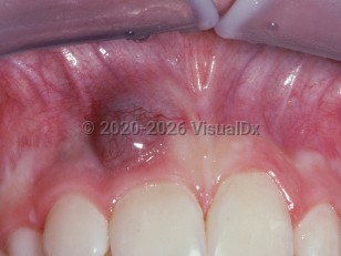
The peripheral giant cell granuloma is a fairly common nodule on the gingiva caused by a proliferation of giant cells. It is likely caused by gingival and/or periodontal inflammation that results in differentiation of pluripotent cells into osteoclast-like or monocyte-like cells.
It tends to occur in older individuals in the fifth and sixth decades with a slight female predilection. The mandibular gingiva is more often involved than the maxillary. It is usually non-painful. Biting on it may induce bleeding since it is contains many blood vessels. It often occurs in the edentulous ridge. The lesion may lie in a cup-shaped depression in the underlying bone. Sometimes it may be difficult to tell if this is a purely extra-osseous process that has cupped the bone or whether it is an intra-osseous central giant cell granuloma that has broken through the bone and now has an extra-osseous or "peripheral" presentation. Unlike the central giant cell granulomas, these are not usually associated with hyperparathyroidism.
Peripheral giant cell granuloma - Oral Mucosal Lesion
Alerts and Notices
Important News & Links
Synopsis

Codes
ICD10CM:
K13.4 – Granuloma and granuloma-like lesions of oral mucosa
SNOMEDCT:
89722009 – Giant cell peripheral granuloma
K13.4 – Granuloma and granuloma-like lesions of oral mucosa
SNOMEDCT:
89722009 – Giant cell peripheral granuloma
Look For
Subscription Required
Diagnostic Pearls
Subscription Required
Differential Diagnosis & Pitfalls

To perform a comparison, select diagnoses from the classic differential
Subscription Required
Best Tests
Subscription Required
Management Pearls
Subscription Required
Therapy
Subscription Required
References
Subscription Required
Last Updated:11/13/2013
Peripheral giant cell granuloma - Oral Mucosal Lesion