Contents

SynopsisCodesLook ForDiagnostic PearlsDifferential Diagnosis & PitfallsBest TestsManagement PearlsTherapyReferences

View all Images (7)

Pigmented contact dermatitis
Other Resources UpToDate PubMed

Pigmented contact dermatitis

Contributors: Alexis E. Carrington MD, Scarlett Boulos MD, Susan Burgin MD
Other Resources UpToDate PubMed

Synopsis

Pigmented contact dermatitis, also known as Riehl melanosis, pigmented cosmetic dermatitis, and melanosis faciei feminae (female facial melanosis), is a nonpruritic pigmented contact dermatitis of rapid onset characterized by reticulated, brownish-gray pigmentation of the face and neck. It particularly affects the forehead, temples, and zygomatic regions.

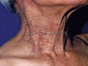
While some aspects of the etiology remain unclear, investigations, particularly in Japan, have found that the hyperpigmentation stems from sensitivity to certain chemicals in cosmetics. In these investigations, hundreds of patients were shown to have positive patch tests to cosmetics and their ingredients, and their hyperpigmentation significantly improved after avoiding cosmetics with those allergens.

Common chemicals implicated in pigmented contact dermatitis include:
  • Fragrances – hydroxycitronellal, benzyl salicylate, jasmine absolute, ylang-ylang oil, cananga oil, sandalwood oil, eugenol, cinnamic derivatives, hydroperoxides of limonene, and balsam of Peru
  • Pigments – D & C Red 31, Red 225; D & C Yellow 11, Yellow 10; and pigments containing phenyl-azo-e-naphthol, aniline dyes, kumkum (a red powder commonly used by Hindu women), and henna
  • Optical whiteners
  • Coal tar derivatives, which increase photosensitivity
  • Bactericidals – carbanilides such as trichlocarban and halocarban
Pigmented cosmetic contact dermatitis most commonly affects women, especially women of Asian and Hispanic descent. Women with very light skin phototypes and very dark skin phototypes do not show the same skin color changes. Men and children can also be affected by pigmented contact dermatitis.

Codes

ICD10CM:
L81.4 – Other melanin hyperpigmentation

SNOMEDCT:
24285001 – Riehl's melanosis

Look For

Subscription Required

Diagnostic Pearls

Subscription Required

Differential Diagnosis & Pitfalls

  • Allergic contact dermatitis – Presents with more prominent signs of erythema, urticaria, vesiculation, and pruritus. Caused by specific allergens; distribution varies and depends on contact.
  • Irritant contact dermatitis – May have erythema, mild edema, and scaling caused by direct contact with chemical agents such as corrosive agents, which can burn and produce ulcers; most commonly affects the hands.
  • Berloque dermatitis (see phytophotodermatitis) – Hyperpigmentation in drop-like or pendant-like configuration in sun-exposed areas where perfume with bergamot oil (UV sensitizer) has been applied.
  • Melasma – Brown-gray hyperpigmentation correlated mostly with sun exposure as well as genetics, pregnancy, hormone replacement therapy, oral contraceptives, and thyroid problems.
  • Exogenous ochronosis – Caused by topical hydroquinone. Erythema and mild dyspigmentation are superseded by brown, blue, and/or black mottled macules.
  • Poikiloderma of Civatte – Common disorder, concerning mainly perimenopausal women; located on the lateral and low neck.
  • Erythromelanosis follicularis of the face and neck – Reddish-brown pigmentation affecting follicles in the periauricular and maxillary areas; pigmented areas blanch with applied pressure, showing more brown pigment and telangiectases. Differing histology on skin biopsy.
  • Addison disease – Hyperpigmentation is more generalized on the body, with other systemic symptoms like hypotension, myalgias, arthralgias, nausea, vomiting or diarrhea, and amenorrhea in women.
  • Lichen amyloidosis – Extremely pruritic eruption of red-brown hyperkeratotic papules, distributed mostly on the shins, thighs, and feet; Congo red stain shows green birefringence with polarizing light.
  • Macular amyloidosis – Symmetric and pruritic eruption of brown or grayish macules located on upper back and arms; same Congo red stain results as for lichen amyloidosis.
  • Hyperpigmented discoid lupus erythematosus – Direct immunofluorescence study on skin biopsy helps differentiate the conditions.

Best Tests

Subscription Required

Management Pearls

Subscription Required

Therapy

Subscription Required

References

Subscription Required

Last Reviewed:09/21/2020
Last Updated:09/21/2020
Copyright © 2023 VisualDx®. All rights reserved.
Pigmented contact dermatitis
A medical illustration showing key findings of Pigmented contact dermatitis : Reticular - netlike, Hyperpigmented macules, Hyperpigmented patches
Clinical image of Pigmented contact dermatitis - imageId=93789. Click to open in gallery.  caption: 'Hyperpigmented and reddish patches on the neck.'
Hyperpigmented and reddish patches on the neck.
Copyright © 2023 VisualDx®. All rights reserved.