Pilar (trichilemmal) cysts, sometimes referred to as wens, are common, benign cysts of hair follicle origin that are most often found on the scalp. They are derived from the outer root sheath of the hair follicle. Histologically, they are lined with multilayered epithelium that does not mature through a granular layer.
The cysts are smooth and mobile, are keratin filled, and may or may not be tender. They occur most commonly in middle-aged individuals, especially women. They may be present as solitary or multiple lesions. Pilar cysts are the most common type of cutaneous cyst of the scalp and the second most common cutaneous cyst of the head and neck (after epidermal inclusion cysts).
Rarely, these cysts form proliferating pilar tumors (more commonly called proliferating trichilemmal cysts), which are benign but may be locally aggressive or ulcerate. Malignant transformation is very rare but can lead to distant metastasis. Verrucous pilar cysts, or pilar cysts with features of human papillomavirus infection on histopathology, have also been described.
Pilar cysts may be inherited as an autosomal dominant trait with incomplete penetrance. Multiple cysts may develop in these cases. A variant of the PLCD1 gene has been shown to be associated with cyst formation in these families, and recent genetic studies suggest that a somatic mutation on the same allele (a monoallelic two-hit mechanism) is the genetic trigger for cyst development.
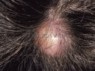
Pilar cyst
See also in: Hair and ScalpAlerts and Notices
Important News & Links
Synopsis

Codes
ICD10CM:
L72.11 – Pilar cyst
SNOMEDCT:
254677004 – Trichilemmal cyst
L72.11 – Pilar cyst
SNOMEDCT:
254677004 – Trichilemmal cyst
Look For
Subscription Required
Diagnostic Pearls
Subscription Required
Differential Diagnosis & Pitfalls

To perform a comparison, select diagnoses from the classic differential
Subscription Required
Best Tests
Subscription Required
Management Pearls
Subscription Required
Therapy
Subscription Required
References
Subscription Required
Last Reviewed:12/12/2019
Last Updated:01/20/2022
Last Updated:01/20/2022
 Patient Information for Pilar cyst
Premium Feature
VisualDx Patient Handouts
Available in the Elite package
- Improve treatment compliance
- Reduce after-hours questions
- Increase patient engagement and satisfaction
- Written in clear, easy-to-understand language. No confusing jargon.
- Available in English and Spanish
- Print out or email directly to your patient
Upgrade Today

Pilar cyst
See also in: Hair and Scalp