Poikiloderma vasculare atrophicans (PVA) is considered by some authors to be a rare variant of early patch-stage mycosis fungoides (MF) and by others to be a precursor to MF. It has also been referred to as poikiloderma atrophicans vasculare or poikiloderma vasculare atrophicans of Jacobi-Lane, or "poikilodermatous" variant of MF.
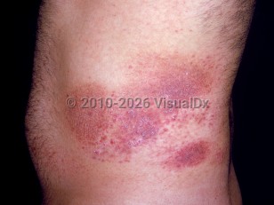
PVA is characterized by atrophic patches with telangiectasia and mottled pigmentation, usually on the trunk and flexural areas. The condition is typically asymptomatic; however, there may be pruritus.
Onset is typically between 40 and 60 years of age, with a slight male predilection. Initial presentation may be with one to a few patches that persist for many years. Over time, the lesions may remain stable in size or gradually enlarge or thicken as well as increase in number. Wrinkled, thin plaques that are not poikilodermatous and that resemble patch-stage MF may also be present.
Progression to a more advanced stage of MF is not reliably predictable, and the disease may remain quiescent for many years or indefinitely.
Poikiloderma vasculare atrophicans
Alerts and Notices
Important News & Links
Synopsis

Codes
ICD10CM:
L94.5 – Poikiloderma vasculare atrophicans
SNOMEDCT:
16341002 – Poikiloderma vasculare atrophicans
L94.5 – Poikiloderma vasculare atrophicans
SNOMEDCT:
16341002 – Poikiloderma vasculare atrophicans
Look For
Subscription Required
Diagnostic Pearls
Subscription Required
Differential Diagnosis & Pitfalls

To perform a comparison, select diagnoses from the classic differential
Subscription Required
Best Tests
Subscription Required
Management Pearls
Subscription Required
Therapy
Subscription Required
References
Subscription Required
Last Reviewed:09/23/2018
Last Updated:10/07/2018
Last Updated:10/07/2018
Poikiloderma vasculare atrophicans