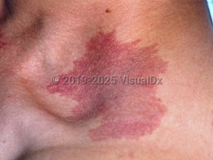
A port-wine stain, or capillary malformation (CM), is a vascular malformation typically found on the face. Lesions are present at birth in 0.3%-0.5% of newborns in the United States, with equal sex distribution. In the past, port-wine stains and salmon patches were considered to be variations of capillary malformation; it is now known that port-wine stains are truly malformations of capillaries that may be associated with systemic complications and will never spontaneously improve. Furthermore, salmon patches represent dilatation of capillaries rather than a true malformation. These often fade with time if located on the face and are not associated with any other abnormalities.
Of facial lesions, about half are restricted to one unilateral segmental region, while the other half are more widespread, cross the midline, or occur bilaterally. Glaucoma occurs in 10% of patients with facial lesions, more often in those that involve the forehead and/or eyelids. When a port-wine stain occurs on the forehead, Sturge-Weber syndrome (encephalotrigeminal angiomatosis – CM of the upper face, ipsilateral leptomeninges, and cerebral cortex) should be considered. This diagnosis is made in less than 10% of patients with upper eyelid or forehead CMs.
Wyburn-Mason syndrome (unilateral retinocephalic syndrome or Bonnet-Dechaume-Blanc syndrome) manifests as CM anywhere on the face (not just periorbital), monocular amblyopia, proptosis, and conjunctival vessel dilation plus or minus facial hypertrophy and central nervous system involvement, with the CM leading to neurologic symptoms.
Phakomatosis pigmentovascularis is a neural crest disorder (usually in those of Asian descent) with a CM in addition to a nevus, usually melanocytic in origin. When oculodermal melanocytosis and CM occur together involving the eye globe, there is a strong predisposition for congenital glaucoma. Choroidal melanoma is another rare complication. See Nevus of Ota for more information.
Lesions persist for life without resolution.
Port-wine stain - External and Internal Eye
See also in: OverviewAlerts and Notices
Important News & Links
Synopsis

Codes
ICD10CM:
Q82.5 – Congenital non-neoplastic nevus
SNOMEDCT:
416377005 – Port-wine stain of skin
Q82.5 – Congenital non-neoplastic nevus
SNOMEDCT:
416377005 – Port-wine stain of skin
Look For
Subscription Required
Diagnostic Pearls
Subscription Required
Differential Diagnosis & Pitfalls

To perform a comparison, select diagnoses from the classic differential
Subscription Required
Best Tests
Subscription Required
Management Pearls
Subscription Required
Therapy
Subscription Required
References
Subscription Required
Last Reviewed:11/16/2017
Last Updated:09/30/2018
Last Updated:09/30/2018
 Patient Information for Port-wine stain - External and Internal Eye
Premium Feature
VisualDx Patient Handouts
Available in the Elite package
- Improve treatment compliance
- Reduce after-hours questions
- Increase patient engagement and satisfaction
- Written in clear, easy-to-understand language. No confusing jargon.
- Available in English and Spanish
- Print out or email directly to your patient
Upgrade Today

Port-wine stain - External and Internal Eye
See also in: Overview