Pressure injuries occur more commonly in certain subsets of patients, such as the elderly, patients who have had surgery for hip fracture, and patients with spinal cord injury. Pressure injuries are also more common in patients who are from nursing homes and assisted living facilities or who are otherwise hospitalized.
Factors promoting pressure injury formation include the following:
- Pressure – The primary contributive factor leading to formation of ulcers. The length of time that high pressures are sustained is just as important as the degree of pressure. Thus, constantly relieving pressure prevents tissue damage or tissue death.
- Friction – Occurs when two surfaces resist movement at their interface, resulting in damage to superficial layers of skin. The epidermis is abraded, and intraepidermal blisters may result that lead to superficial skin erosions, not unlike a mild burn. This can occur when a patient is dragged across a bed sheet (referred to as a sheet burn) or when a patient wears a badly fitting prosthetic device.
- Shearing Forces – Generated by the motion of bone and subcutaneous tissue relative to the skin. Gravity acts to pull the body down, which is prevented from moving due to friction (as seen when the head of the bed is raised to more than 30 degrees or when a seated patient slides down a chair). Even though the body is being pulled down or forward, the skin is pinned in its place. This involves the interplay of gravity and friction to propagate these shearing forces. Shear is responsible for a large amount of the damage associated with pressure injuries.
- Moisture – Moist surfaces predispose to ulcer formation in two ways: First, moisture increases the effects of pressure, friction, and shear on the skin by decreasing its resistance to these forces and secondly by causing maceration of the skin, thereby increasing the incidence of ulcer formation fivefold. Conditions leading to excess moisture may arise due to perspiration, urinary or fecal incontinence, or leakage from a wound site.
- Stage 1: Skin is intact with an area of non-blanching erythema. This is usually over a bony prominence.
- Stage 2: Partial thickness skin loss with loss of the epidermis and some of the dermis. It appears as a shallow ulcer with a red-pink color. No slough or necrotic tissue is present in the base. It may also appear as an enclosed or open serum-filled blister.
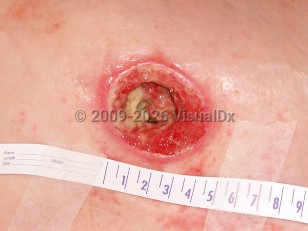
- Stage 3: Full thickness loss of skin with the epidermis and dermis gone and damage to or necrosis of subcutaneous tissues. Damage extends down to but not through the underlying fascia. Subcutaneous fat may be visible, but muscle, tendon, or bone is not seen. Slough may be present but does not hinder estimation of the extent of tissue loss. Tunneling or undermining may be present.
- Stage 4: Full thickness loss of skin with extensive destruction, tissue necrosis, and damage to bone, muscle, or other supporting structures that are exposed.
- Deep tissue pressure injury: Area of localized, discolored intact skin that is purple or maroon-red in color. It may also appear as a blood-filled blister resulting from damage to underlying soft tissue. Preceding skin changes may include skin that is painful, firm, boggy, or that has a different temperature as compared to the surrounding skin.
- Unstageable: Full tissue thickness loss in which the base of the ulcer is covered by slough or an eschar and, therefore, the true depth of the damage cannot be estimated until these are removed.
- Medical device-related pressure injury (describes an etiology): Results from the use of devices designed and applied for therapeutic purposes. Injury generally conforms to the pattern or shape of the device. Stage using the staging system.
- Mucosal membrane pressure injury: Found on mucous membranes with a history of a medical device in use at the location of the injury. Cannot be staged due to anatomy of tissue.
Risk factors leading to pressure injury formation are as follows:
- Limited mobility
- Malnutrition
- Anemia
- Advanced age
- Fecal or urinary incontinence
- Smoking
- Dry skin
- Altered skin perfusion – Decreased in cases of shock or increased if patient has fluid edema due to overhydration.
- Acute illness leading to temporary immobility
- Chronic systemic illness
- Terminal illness
- Degenerative neurologic disease
- Increased weight
- Sudden decrease in weight
- Altered mental status
- Prolonged pressure
- Corticosteroids
- Sedatives
- Analgesics
- Anti-hypertensives
- Assess and record the stage of the ulcer and the location according to ICD codes.
- Carry out an assessment using the Braden or Norton scale. These act as tools for predicting pressure injury risk; this should be done for patients who have not yet developed an ulcer but could be susceptible to one and those who have already developed one. This is an important assessment, as it determines the prevention measures taken and the type of pressure-reducing support surfaces consequently used.
- Monitor the progress daily.

 Patient Information for