David Adelson MD
Christine Ahn MD
Sanah Ali MD
Carl Allen DDS, MSD
Brandon Ayres MD
Howard P. Baden MD
Yevgeniy Balagula MD
Robert Baran MD
Keira Barr MD
Sid Barsky MD
Gregory J. Basura MD, Ph.D
Donald Belsito MD
Jeffrey D. Bernhard MD
Jesse Berry MD
Victor Blanco MD
Benjamin R. Bohaty MD
William Bonnez MD
Mina Botros MD
Jeremy S. Boyd MD
Sarah Brenner MD
Robert A. Briggaman MD
Robert Brodell MD
Roman Bronfenbrener MD
Walter Brooks MD
Daniel Bryan MD
William Buckley MD
Philip Bulterys MD, PhD
Susan Burgin MD
Sonya Burton MD
Sean P. Bush MD, FACEP
Jeffrey Callen MD
Scott Camazine MD
Michael Cardwell
Shelley D. Cathcart MD
Robert Chalmers MD, MRCP, FRCP
Chia-Yu Chu MD, PhD
Flavio Ciferri MD
Maria Rosa Cordisco MD
Noah Craft MD, PhD
John T. Crissey MD
Harold E. Cross MD, PhD
Charles E. Crutchfield III MD
Adriana Cruz MD
Donna Culton MD, PhD
Bart J. Currie MBBS, FRACP, DTM&H
Chicky Dadlani MD
Alexander Dane DO
Roxana Daneshjou MD
C. Ralph Daniel III MD
Thomas Darling MD, PhD
Steven Daveluy MD
William Delaney MD
Shreya Deoghare MD
Alex Derstenfeld
Damian P. DiCostanzo MD
Benedict F. DiGiovanni MD
Ncoza Dlova MD
Erin Doudt
James Earls MD
Libby Edwards MD
Melissa K. Egge MD
Charles N. Ellis MD
Rachel Ellis MD
David Elpern MD
Nancy Esterly MD
Stephen Estes MD
E. Dale Everett MD
Janet Fairley MD
David Feingold MD
Jennifer J. Findeis-Hosey MD
Benjamin Fisher MD
Robert Warne Fitch, MD
Henry Foong MBBS, FRCP
David Foster MD, MPH
Lindy P. Fox MD
Brian D. Foy PhD
Michael Franzblau MD
Vincent Fulginiti MD
Sunir J. Garg MD, FACS
Kevin J. Geary MD
Sharon Glick MD
Lowell Goldsmith MD, MPH
Sethuraman Gomathy MD
Bernardo Gontijo MD, PhD
Ayman Grada MD
Kenneth Greer MD
Kenneth G. Gross MD
Alan Gruber MD
Nathan D. Gundacker MD
Akshya Gupta MD
Vidal Haddad MSC, PhD, MD
Edward Halperin MD, MA
Ronald Hansen MD
John Harvey
Rizwan Hassan MD
Michael Hawke MD
Jason E. Hawkes MD
Peter W. Heald MD
Martha Hickman MD
David G. Hicks MD
Sarah Hocker DO
Ryan J. Hoefen MD, PhD
Li-Yang Hsu MD
Jennifer Huang MD
William W. Huang MD, MPH, FAAD
Eric F. Ingerowski MD
Sanjana Iyengar MD
Alvin H. Jacobs MD
Randy Jacobs MD, FAAD
Saagar Jadeja MD
Shahbaz A. Janjua MD
Joshua J. Jarvis MD
Noman Javed, MD
Beverly A. Johnson MD
Kit Johnson
Zachary John Jones MD
Harsimran Kaur MD
Robert Kalb MD
Katherine Kaproth-Joslin MD, PhD
Philip J. Katzman MD
A. Paul Kelly MD
Henry Kempe MD
Loren Ketai MD
Sidney Klaus MD
Ashwin Kosambia MD
Jessica A. Kozel MD
Carl Krucke
Mario E. Lacouture MD
Joseph Lam MD
Alfred T. Lane MD
Gerald S. Lazarus MD
Edith Lederman MD
Nahyoung Grace Lee MD
Ronald J. Lee MD
Pedro Legua MD, PhD
Robert Levin MD
Bethany Lewis MD
Sue Lewis-Jones FRCP, FRCPCH
Taisheng Li MD
Christine Liang MD
Shari Lipner MD, PhD
Adam Lipworth MD
Jenna Lullo MD
Jason Maguire MD
Mark Malek MD, MPH
Jere Mammino DO
Ricardo Mandojana MD
Lynne Margesson MD
Thomas J. Marrie MD
Maydel Martinez MD
Ralph Massey MD
Patrick McCleskey MD
Lynn McKinley-Grant MD
Karen McKoy MD
Thomas McMeekin MD
Josette McMichael MD
Somchai Meesiri MD
Joseph F. Merola MD
Mary Gail Mercurio MD
Anis Miladi MD
Larry E. Millikan MD
Dan Milner Jr. MD
Zaw Min MD
Nequesha Mohamed MD
Stephanie Montero
Alastair Moore MD
Martha Alejandra Morales-Sánchez MD
Keith Morley MD
Dean Morrell MD
Samuel Moschella MD
Surya N. Mundluru MD
Nicholas Erick Nacca MD
Rehan Naseemuddin MD
Taimor Nawaz MD
Vic Newcomer MD
John Nguyen MD
Matilda Nicholas MD, PhD
Thomas P. Nigra MD
Dylan Norton MD
Steven Oberlender MD, PhD
Maria Teresa Ochoa MD
Edward O'Keefe ND
Daniel C. Oppenheimer MD
Art Papier MD
Lawrence Parish MD
Tanner Parrent MD
Mukesh Patel MD
Lauren Patty-Daskivich MD
David Peng MD, MPH
Robert Penne MD
Nitipong Permpalung MD
Frank Johannes Plate MD, PhD
Miriam Pomeranz MD
Doug Powell MD
Harold S. Rabinovitz MD
Christopher J. Rapuano MD
Sireesha Reddy MD
Angela Restrepo MD, PhD
Cynthia Reyes-Barron MD
Bertrand Richert MD, PhD
J. Martin Rodriguez, MD, FACP
Theodore Rosen MD
Misha Rosenbach MD
Scott Schiffman MD
Robert H. Schosser MD
Glynis A. Scott MD
Carlos Seas MD, MSc
Deniz Seçkin MD
Daniel Sexton MD
Paul K. Shitabata MD
Tor Shwayder MD, FAAP, FAAD
Daniel Siegel, MD
Elaine Siegfried MD
Gene Sienkiewicz MD
Christye Sisson
Jonathan Soh MD
Philip I. Song MD
Mary J. Spencer MD, FAAP
Lawrence B. Stack MD
Sarah Stein MD
William Van Stoecker MD
Frances J. Storrs MD
Erik J. Stratman MD
Lindsay C. Strowd MD
Erika Summers MD
Belinda Tan MD, PhD
Robert Tomsick MD
Hensin Tsao MD, PhD
Richard P. Usatine MD
Jenny Valverde MD
Vishalakshi Viswanath MD
Susan Voci MD
Lisa Wallin ANP, FCCWS
Douglas Walsh MD
Ryan R. Walsh MD
George Watt MD
Erin X. Wei MD
Zachary Werle DO
Clayton E. Wheeler MD
Sally-Ann Whelan MS, NP, CWOCN
Danielle Wilbur MD
Jan Willems MD, PhD
Victoria L. Williams MD
James Henry Willig MD, MPH
Kelly Wilmas MD
Karen Wiss MD
Vivian Wong MD, PhD
Sook-Bin Woo MS, DMD, MMSc
Jamie Woodcock MD
Stephen J. Xenias MD
Nathaniel Yohannes
Lisa Zaba MD
Vijay Zawar MD
Bonnnie Zhang MD
Carolyn Ziemer MD
Barret Zimmerman MD
Jeffrey P. Zwerner MD, PhD
Organizations
American Academy of Dermatology
Am. Journal of Trop. Med & Hygiene
Armed Forces Pest Management Board
Blackwell Publishing
Broward Health Medical Center
Bugwood Network
Centers For Disease Control and Prevention
Centro Internacional de Entrenamiento e Investigaciones Mèdicas (CIDEIM)
Dermatology Associates of Concord
Dermatology Online Journal
East Carolina University (ECU), Division of Dermatology
International Atomic Energy Agency
Massachusetts Medical Society
NYU Department of Dermatology
Oregon Health & Science University (OHSU)
Oxford University Press
Radiological Society of North America
Washington Hospital Center
Wikipedia
World Health Organization
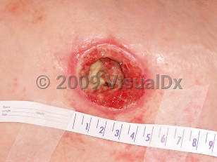
A pressure injury results when there is localized damage to the skin and underlying tissue as a result of compression between a bony prominence and an external surface. Damage is caused by the forces of pressure, shear, and friction acting individually or in combination with each other. Pressure injuries, previously termed as decubitus ulcers, are also commonly referred to as pressure sores and bed sores. Common sites for pressure injury formation are the sacrum, over the ischial tuberosity, the trochanter, and the calcaneus. Other locations are the elbow, ankle, scapula, and the occiput. However, the most common sites are the sacrum and the heels. Pressure injuries affect from 1.5-3 million people in the US, at an annual cost of approximately 5 billion dollars.
Pressure injuries occur more commonly in certain subsets of patients, such as the elderly, patients who have had surgery for hip fracture, and patients with spinal cord injury. Pressure injuries are also more common in patients who are from nursing homes and assisted living facilities or who are otherwise hospitalized.
Factors promoting pressure injury formation include the following:
Pressure – The primary contributive factor leading to formation of ulcers. The length of time that high pressures are sustained is just as important as the degree of pressure. Thus, constantly relieving pressure prevents tissue damage or tissue death.
Friction – Occurs when two surfaces resist movement at their interface, resulting in damage to superficial layers of skin. The epidermis is abraded, and intraepidermal blisters may result that lead to superficial skin erosions, not unlike a mild burn. This can occur when a patient is dragged across a bed sheet (referred to as a sheet burn) or when a patient wears a badly fitting prosthetic device.
Shearing Forces – Generated by the motion of bone and subcutaneous tissue relative to the skin. Gravity acts to pull the body down, which is prevented from moving due to friction (as seen when the head of the bed is raised to more than 30 degrees or when a seated patient slides down a chair). Even though the body is being pulled down or forward, the skin is pinned in its place. This involves the interplay of gravity and friction to propagate these shearing forces. Shear is responsible for a large amount of the damage associated with pressure injuries.
Moisture – Moist surfaces predispose to ulcer formation in two ways: First, moisture increases the effects of pressure, friction, and shear on the skin by decreasing its resistance to these forces and secondly by causing maceration of the skin, thereby increasing the incidence of ulcer formation fivefold. Conditions leading to excess moisture may arise due to perspiration, urinary or fecal incontinence, or leakage from a wound site.
Pressure injuries are classified according to the extent of tissue damage, per the National Pressure Ulcer Advisory Panel (NPUAP):
Stage 1: Skin is intact with an area of non-blanching erythema. This is usually over a bony prominence.
Stage 2: Partial thickness skin loss with loss of the epidermis and some of the dermis. It appears as a shallow ulcer with a red-pink color. No slough or necrotic tissue is present in the base. It may also appear as an enclosed or open serum-filled blister.
Stage 3: Full thickness loss of skin with the epidermis and dermis gone and damage to or necrosis of subcutaneous tissues. Damage extends down to but not through the underlying fascia. Subcutaneous fat may be visible, but muscle, tendon, or bone is not seen. Slough may be present but does not hinder estimation of the extent of tissue loss. Tunneling or undermining may be present.
Stage 4: Full thickness loss of skin with extensive destruction, tissue necrosis, and damage to bone, muscle, or other supporting structures that are exposed.
Deep tissue pressure injury: Area of localized, discolored intact skin that is purple or maroon-red in color. It may also appear as a blood-filled blister resulting from damage to underlying soft tissue. Preceding skin changes may include skin that is painful, firm, boggy, or that has a different temperature as compared to the surrounding skin.
Unstageable: Full tissue thickness loss in which the base of the ulcer is covered by slough or an eschar and, therefore, the true depth of the damage cannot be estimated until these are removed.
The NPUAP 2016 updated staging system also includes the following:
Medical device-related pressure injury (describes an etiology): Results from the use of devices designed and applied for therapeutic purposes. Injury generally conforms to the pattern or shape of the device. Stage using the staging system.
Mucosal membrane pressure injury: Found on mucous membranes with a history of a medical device in use at the location of the injury. Cannot be staged due to anatomy of tissue.
When estimating the depth of pressure injuries for purposes of staging, it is important to keep in mind anatomical location, especially in the case of stage 3 ulcers. Ulcers on the bridge of the nose, ear, occiput, and malleolus do not have subcutaneous tissue and, consequently, will be very shallow (but still graded as stage 3), as compared to a stage 3 ulcer on the sacrum.
Risk factors leading to pressure injury formation are as follows:
Limited mobility
Malnutrition
Anemia
Advanced age
Fecal or urinary incontinence
Smoking
Dry skin
Altered skin perfusion – Decreased in cases of shock or increased if patient has fluid edema due to overhydration.
Acute illness leading to temporary immobility
Chronic systemic illness
Terminal illness
Degenerative neurologic disease
Increased weight
Sudden decrease in weight
Altered mental status
Prolonged pressure
Simultaneous treatment with the following medications can also predispose to ulcer formation:
Corticosteroids
Sedatives
Analgesics
Anti-hypertensives
When a patient presents with a pressure injury, the following criteria should initially be followed:
Assess and record the stage of the ulcer and the location according to ICD codes.
Carry out an assessment using the Braden or Norton scale. These act as tools for predicting pressure injury risk; this should be done for patients who have not yet developed an ulcer but could be susceptible to one and those who have already developed one. This is an important assessment, as it determines the prevention measures taken and the type of pressure-reducing support surfaces consequently used.
Monitor the progress daily.
ICD codes identifying both the site and stage of the ulcer are needed for complete staging of the pressure injury.
Codes
ICD10CM: L89.90 – Pressure ulcer of unspecified site, unspecified stage L89.91 – Pressure ulcer of unspecified site, stage 1 L89.92 – Pressure ulcer of unspecified site, stage 2 L89.93 – Pressure ulcer of unspecified site, stage 3 L89.94 – Pressure ulcer of unspecified site, stage 4