Contents

SynopsisCodesLook ForDiagnostic PearlsDifferential Diagnosis & PitfallsBest TestsManagement PearlsTherapyReferences

View all Images (9)

Restrictive dermopathy
Other Resources UpToDate PubMed

Restrictive dermopathy

Contributors: Preethi Ramaswamy MD, Amit Garg MD, Jeffrey D. Bernhard MD, Craig N. Burkhart MD, Dean Morrell MD, Lowell A. Goldsmith MD, MPH
Other Resources UpToDate PubMed

Synopsis

Restrictive dermopathy is a rare lethal laminopathy inherited in an autosomal recessive fashion. A mutation in the zinc metalloproteinase (ZMPSTE24) gene or lamin A is responsible for the condition. Both sexes can be affected. Parental consanguinity increases risk.

Lamins are nuclear proteins that form a network of filaments beneath the nuclear membrane to maintain membrane integrity and control gene expression. Post-translation modification of prelamin A through proteolytic cleavage by zinc metalloproteinase enzyme (ZMPSTE24) results in the formation of mature lamin A. A defect in the enzyme (majority of cases) or in lamin A can lead to the clinical features of restrictive dermopathy.

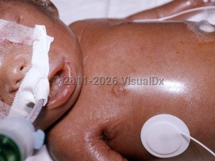
Decrease in intrauterine growth, restricted fetal movement, and polyhydramnios result in premature rupture of membranes and delivery of the child by 31 weeks of gestation. Typical clinical features at birth include tight, rigid, translucent skin; prominent superficial vessels; fixed, open mouth with retromicrognathism; small, pinched nose; multiple joint contractures; and erosions at flexure or pressure sites. Neonates with restrictive dermopathy die within few hours to a week after birth, usually because of respiratory insufficiency secondary to pulmonary hypoplasia.

Codes

ICD10CM:
Q82.8 – Other specified congenital malformations of skin

SNOMEDCT:
400128006 – Lethal tight skin contracture syndrome

Look For

Subscription Required

Diagnostic Pearls

Subscription Required

Differential Diagnosis & Pitfalls

  • Sclerema neonatorum – Occurs in severely ill premature neonates in the first week of life. Characterized by diffuse, cold, board-like hardening of the skin with respiratory difficulties, congestive heart failure, diarrhea, and sepsis. Precipitating factors include hypothermia, perinatal asphyxia, dehydration, and defective complement activity. Can be fatal.
  • Infantile systemic hyalinosis – Autosomal recessive disorder with mutations in the capillary morphogenesis protein-2 (CMG2). Clinical features include diffusely thickened skin with firm papules and nodules on the ears and perinasal and perianal areas. Other features include joint contractures, diarrhea, recurrent infections, and visceral involvement, with hyaline deposits in multiple organs, resulting in early death during infancy.
  • Stiff skin syndrome – Occurs in infancy or early childhood with rock-hard skin bound to underlying tissues, most prominently in areas with fascia such as the buttocks and thighs. Overlying skin may have hypertrichosis and mild hyperpigmentation. Other features include joint contractures and scoliosis. The disease is slowly progressive and nonfatal.
  • Winchester syndrome – Autosomal recessive disorder with mutations in matrix metalloproteinase-2. Clinical features include short stature, progressive articular contractures, corneal opacities, thickened skin with hyperpigmentation, and coarsened facial features. The disease is progressive and ultimately leads to disability.
  • Hutchinson-Gilford progeria syndrome (HGPS) – Neonates are healthy appearing at birth; however, around 18-24 months, growth curves fall below average and characteristic facial features develop, including large head in relation to the body; small, pinched nose; narrow face; alopecia; and scleroderma-like tightening of the skin. A point mutation in the lamin A gene is responsible for this condition. Accumulation of altered lamin A and disruption of nuclear membrane integrity are thought to be responsible for the premature aging process. Atherosclerosis leads to premature death, usually in the teenage years.
  • Familial partial lipodystrophy of Dunnigan variety (FPLD) – Autosomal dominant disorder due to mutation in the lamin A gene. Characterized by gradual loss of adipose tissue from extremities and trunk and excess fat accumulation on the face and neck at puberty. Others features include short stature, scleroderma-like skin, and early graying of hair.
  • Emery-Dreifuss muscular dystrophy (EDMD) – Rare X-linked form of muscular dystrophy due to mutation in the emerin (EMD) gene. The dominant and autosomal forms are due to mutations in the lamin A gene. Characterized by difficulty walking due to muscle wasting and weakness, scoliosis of the spine, and contractures of the Achilles tendon.
  • Mandibuloacral dysplasia, type B – Clinical features include a progeroid appearance, lipodystrophy, mandibular and clavicular hypoplasia, joint contractures, and skin atrophy with mottled pigmentation. There are few case reports of patients with compound heterozygous mutations in the zinc metalloproteinase gene responsible for this condition.

Best Tests

Subscription Required

Management Pearls

Subscription Required

Therapy

Subscription Required

References

Subscription Required

Last Updated:02/03/2022
Copyright © 2023 VisualDx®. All rights reserved.
Restrictive dermopathy
A medical illustration showing key findings of Restrictive dermopathy : Erythema, Flexural distribution, Joint contractures, Micrognathia, Skin erosion, Taut, shiny skin, Ectropion, Hardened skin
Clinical image of Restrictive dermopathy - imageId=5455550. Click to open in gallery.  caption: 'Diffuse shiny, bound-down skin with linear erosions near the axilla.'
Diffuse shiny, bound-down skin with linear erosions near the axilla.
Copyright © 2023 VisualDx®. All rights reserved.