In the United States and other developed countries, the incidence of rheumatic fever is 0.1-2 cases per 100 000 persons. However, the incidence is much higher (about 10-20 cases per 100 000 persons) in developing countries of the world. The lower incidence in developed countries is due to improved public health and living conditions, the use of antibiotics, and a shift in the endemic strains of GAS. The peak onset is between the ages of 6 and 20 years. It affects both sexes equally. Populations at increased risk for streptococcal pharyngitis include military recruits, persons in close contact with school-aged children, and individuals of low socioeconomic status.
The disease manifestations of ARF usually follow streptococcal pharyngitis by 2-3 weeks. It is a clinical diagnosis, and multiple organ systems may be involved. There are 5 major clinical manifestations of ARF, and they are, in order of frequency:
- Polyarthritis (75%),
- Carditis with valvular involvement (40%-50%),
- Sydenham chorea (15%),
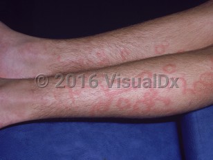
- Erythema marginatum (< 10%), and
- Subcutaneous nodules (< 10%).
Cardiac involvement may include pancarditis involving the pericardium, epicardium, myocardium, and endocardium. The most characteristic component of carditis is valvulitis, with isolated mitral valve disease being the most common development (60% of patients) followed by combined mitral and aortic valve disease; the pulmonic valve is infrequently involved. Valvular disease typically starts with valve dilatation followed by regurgitation. Chronic valvular regurgitation may progress to valvular stenosis. Carditis presents clinically as new murmur, tachycardia, rhythm disturbance, pericardial friction rub, or heart failure. There are 3 distinct murmurs of acute rheumatic carditis depending on the affected cardiac valves, namely:
- A high-pitched blowing pansystolic apical murmur of mitral regurgitation,
- A low-pitched mid-diastolic apical flow murmur of mitral stenosis (Carey Coombs murmur), and
- A high-pitched decrescendo diastolic murmur of aortic regurgitation.
Dermatological manifestations include erythema marginatum and subcutaneous nodules, although these are infrequent (present in < 10% of cases). Erythema marginatum is an evanescent, non-pruritic, non-painful, blanching erythematous macular rash with a pale center, usually affecting the trunk and sometimes the proximal parts of the limbs but not the face. Its presence is suggestive of coexisting carditis. Subcutaneous nodules are firm, painless, and freely mobile. They are found on the extensor surfaces of joints (knees, elbows, wrists, and ankles), bony prominences, tendons, the dorsum of foot, the occipital region, and cervical processes. Patients typically present with 3-4 nodules ranging in size from a few millimeters to 1-2 cm.
Other clinical manifestations include fever, arthralgia, epistaxis, abdominal pain, rheumatic pneumonia, hematuria, and encephalitis.
Pediatric patient considerations: Rheumatic fever is principally a disease of childhood; however, it is rare before the age of 5 years because a mature immune system is required in the pathogenesis of this disease. Joint involvement is less severe in children compared with teenagers and adults.