Rhodococcus equi is an obligate aerobic, asporogenous, non-motile, gram-positive rod (coryneform bacteria) that can appear bacillary in liquid media and coccoid on solid media. It is named after the salmon-pink to red mucoid colonies (red coccus) that it forms on solid media and was originally thought to be solely a veterinary pathogen primarily found in foals. It was not described in humans until 1967, when it was seen in an immunocompromised patient who presented with fever and a cavitary lung lesion.
It most commonly (85% of reported cases) infects patients who are immunocompromised (having impaired cell-mediated immunity). HIV-infected patients account for two-thirds of cases. Other reported associations include organ transplant (most commonly renal transplant), diabetes, alcohol use disorder, chronic renal failure, leukemia, lymphoma, lung cancer, sarcoidosis, chemotherapy, corticosteroid use, and treatment with monoclonal antibodies. There have been fewer documented cases of R. equi infection in patients with HIV in recent years, largely due to antiretroviral therapy and possibly prophylaxis with azithromycin.
Rhodococcus equi has been isolated from water and soil worldwide and is found where livestock defecate (herbivore manure). It is primarily transmitted through inhalation of dust particles during summer seasons in temperate climates but can also be transmitted through ingestion and direct inoculation. However, there have been published cases of immunocompromised patients who denied such exposure yet were still found to have the disease.
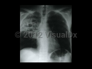
Typically, patients present with pulmonary disease with a subacute course. Main complaints include cough (productive or non-productive), fatigue, fever, and sometimes pleuritic chest pain. Hemoptysis has been reported in 15% of cases. Rhodococcus equi bacteremia frequently complicates pneumonia. Other complications may occur: lung abscess, endobronchial lesions, pleural effusion, empyema, pericarditis, cardiac tamponade, and mediastinitis. Upper lobe cavitary and/or nodular disease is found on radiography, leading to frequent misdiagnosis of tuberculosis. This misdiagnosis can be further compounded by the occasional acid-fast positive stain of the organism. Infection in other locations is usually a late manifestation of pulmonary infection.
Related topic: community-acquired pneumonia
Rhodococcus equi infection
Alerts and Notices
Important News & Links
Synopsis

Codes
ICD10CM:
A49.9 – Bacterial infection, unspecified
SNOMEDCT:
698227004 – Infection due to Rhodococcus equi
A49.9 – Bacterial infection, unspecified
SNOMEDCT:
698227004 – Infection due to Rhodococcus equi
Look For
Subscription Required
Diagnostic Pearls
Subscription Required
Differential Diagnosis & Pitfalls

To perform a comparison, select diagnoses from the classic differential
Subscription Required
Best Tests
Subscription Required
Management Pearls
Subscription Required
Therapy
Subscription Required
Drug Reaction Data
Subscription Required
References
Subscription Required
Last Updated:12/02/2020