- Assess the patient's airway, breathing, circulation, and hemodynamic stability.
- Rule out life-threatening emergencies such as tension pneumothorax, cardiac tamponade, or cardiogenic / hypovolemic shock.
- Determine the mechanism of the patient's injury.
- Complete a secondary survey of the patient to evaluate for additional injuries.
- Obtain a chest x-ray to evaluate the location and severity of the patient's fractures.
- Provide initial pain relief with acetaminophen or NSAIDs and advance as appropriate.
- In the event of major trauma, obtain basic laboratory tests and imaging.
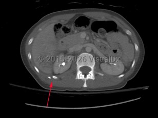
Causes / typical injury mechanism: Rib fractures are a common injury of the thoracic cage. They may be traumatic or pathologic. Depending on the location and the number of ribs involved, be alert for possible visceral injuries, pulmonary contusions, pneumothorax, or vascular injury.
Classic history and presentation: The patient will likely have sustained blunt force trauma to the chest. Common signs and symptoms include chest tenderness or pain, decreased breath sounds, bony crepitus, pain on inspiration, dyspnea, and tachypnea.
In children, certain fractures should increase suspicion for nonaccidental trauma, including rib fractures, often in the context of a delay to seek medical care or inconsistencies in patient history.
Significant rib tenderness and pain without an obvious traumatic cause may be due to a stress fracture from repetitive motions (rowers typically in the 1-3 ribs) or a pathologic fracture from malignancy.
Depending on the location and number of fractured ribs, treatments include body repositioning, medication for pain and inflammation control, ventilation, and incentive spirometry.
Older adults and those with underlying cardiopulmonary disease require close monitoring for complications due to secretions, including atelectasis, hypoventilation, and pneumonia, as older adults have a 5 times higher risk of mortality with the development of pneumonia in the presence of rib fractures.
Risk factors:
- Blunt or penetrating trauma to the chest
- Motor vehicle accident
- Falls from heights greater than 10 feet
- Gunshot wounds to the chest
- Contact sports injury
- Other high-velocity mechanism injuries
- Repetitive motions leading to stress fractures
- Bony neoplasm in rib
Due to their proximity to the subclavian vessels and brachial plexus, upper rib fractures (ribs 1 and 2) can lead to neurovascular injury.
Fractures of the lower ribs (9-12) could indicate concomitant intraabdominal injury.
Grade / classification system:
Rib fractures are commonly described as:
- Rib number(s), right or left
- Acute or chronic with evidence of healing from a remote incident
- Nondisplaced, > 2 mm displacement, comminuted, or flail chest
- Presence or absence of visceral injury
- Age (1 point for every decade)
- Number of rib fractures (3 points per rib)
- Chronic lung disease (5 points)
- Pre-injury anticoagulant use (4 points)
- Oxygen saturation (2 points for every 5% decrease)
- 0-10 points = 13% +/- 6%
- 11-15 points = 29% +/- 8%
- 16-20 points = 52% +/- 8%
- 21-25 points = 70% +/- 6%
- 26-30 points = 80% +/- 6%
- 31+ points = 88% +/- 7%