Contents

SynopsisCodesLook ForDiagnostic PearlsDifferential Diagnosis & PitfallsBest TestsManagement PearlsTherapyReferences

View all Images (6)

Sneddon syndrome in Child
Other Resources UpToDate PubMed

Sneddon syndrome in Child

Contributors: Craig N. Burkhart MD, Dean Morrell MD, Lowell A. Goldsmith MD, MPH
Other Resources UpToDate PubMed

Synopsis

In children, Sneddon syndrome, also known as idiopathic livedo reticularis with cerebrovascular accidents, is a very rare (and likely underdiagnosed) syndrome that consists of ischemic strokes and generalized livedo racemosa. Less than a dozen cases are reported in children and there may be confusion between Sneddon syndrome, cutis marmorata telangiectatica congenita, and Moyamoya syndrome. Knowing which individuals with persistent livedo reticularis are most prone to cerebral vascular occlusion would be very important for affected individuals, but this is not yet possible.

The key cutaneous feature is a persistent livedo reticularis, often in a livedo racemosa pattern (a form of livedo reticularis consisting of a large branching pattern, usually on the trunk, limbs, and proximal extremities). Individuals with Sneddon syndrome, including children, may have antiphospholipid antibodies. The prevalence has been reported to be from 0% to 85%. Regardless of whether patients have antiphospholipid antibodies, individuals with Sneddon syndrome have occlusive arteriopathy and endothelial damage. Up to 35% will have antiendothelial cell antibodies.

Clinical features:
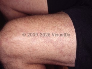
  • Livedo racemosa – Often precedes the onset of focal neurologic symptoms.
  • Headache and vertigo – Most frequent symptoms; may precede livedo racemosa by several years but are nonspecific.
  • Ischemic strokes – Most symptoms are from middle cerebral artery infarcts and thalamic infarcts: hemiparesis, sensory alterations, and aphasia. Less commonly, there may be dysarthria, visual field deficits, or sudden spontaneous falls.
  • Transient ischemic attacks
  • Vascular dementia
  • Secondary symptoms and other organ manifestations in children include growth and intellectual disability.
  • Moyamoya disease – In childhood, presents with ischemic attacks or strokes with occluded intra- and extracerebral arteries. Skin manifestations are not common symptoms of the disease.

Codes

ICD10CM:
M30.8 – Other conditions related to polyarteritis nodosa

SNOMEDCT:
238776001 – Sneddon syndrome

Look For

Subscription Required

Diagnostic Pearls

Subscription Required

Differential Diagnosis & Pitfalls

Many other causes of persistent livedo reticularis or livedo racemosa are in the differential diagnosis, particularly because cutaneous findings in Sneddon syndrome will often precede neurological symptoms.
  • Cutis marmorata telangiectasia congenita – Present at birth, often limited to a limb that is hypotrophic, and may have ulceration.
  • Livedoid vasculopathy – Painful punched-out ulcers around ankles; tends to affect middle-age and elderly individuals.
  • Polyarteritis nodosa – Cutaneous variant (more common in children) is more likely to have livedo racemosa and nodules. Histology: leukocytoclastic vasculitis.
  • Cholesterol emboli or atrial myxoma emboli – Characteristic histology shows cholesterol clefts within thrombi with cholesterol emboli.
  • Necrotizing livedo reticularis – May involve cutaneous nodules and ulcerations. This has many disease associations, including leukocytoclastic vasculitis, calciphylaxis, cryoglobulinemia, granulomatosis with polyangiitis, atrial myxomas, Graves disease, connective tissue diseases, acute lymphocytic leukemia, and renal cancer.
  • Amantadine-induced livedo reticularis – Usually occurs on the legs.
  • Primary antiphospholipid antibody syndrome with livedo reticularis is associated with epilepsy.
  • Systemic lupus erythematosus (SLE) with livedo reticularis is associated with more severe SLE.
  • Essential thrombocythemia
  • Thromboangiitis obliterans – Rest pain, chronic ulcers on distal extremities, gangrene of fingers and/or toes.
  • Moyamoya syndrome – Shows stenosis or occlusion of extracranial and intracranial blood vessels.
  • Malignant atrophic papulosis (Degos disease) – May exhibit some clinical features that overlap with those of Sneddon syndrome (eg, cerebrovascular accident in a young person without other known risk factors).

Best Tests

Subscription Required

Management Pearls

Subscription Required

Therapy

Subscription Required

References

Subscription Required

Last Updated:09/30/2018
Copyright © 2023 VisualDx®. All rights reserved.
Sneddon syndrome in Child
A medical illustration showing key findings of Sneddon syndrome : Headache, Antiphospholipid antibodies present, Aphasia, Trunk, Vertigo, Visual field defect, BP increased, Arms, Legs
Clinical image of Sneddon syndrome - imageId=4342337. Click to open in gallery.  caption: 'Dusky retiform patches (livedo racemosa) on the thigh.'
Dusky retiform patches (livedo racemosa) on the thigh.
Copyright © 2023 VisualDx®. All rights reserved.