Contents

SynopsisCodesLook ForDiagnostic PearlsDifferential Diagnosis & PitfallsBest TestsManagement PearlsTherapyDrug Reaction DataReferences

View all Images (2)

Solar urticaria in Infant/Neonate
Other Resources UpToDate PubMed

Solar urticaria in Infant/Neonate

Contributors: Elyse M. Love MD, Susan Burgin MD, Keith Morley MD
Other Resources UpToDate PubMed

Synopsis

Solar urticaria is a rare, intermittent cutaneous eruption characterized by urticaria induced by ultraviolet and/or visible light. The inducible action spectrum varies by individual and may evolve with time but typically includes the ultraviolet A (UVA) and visible light spectrum; rarely, ultraviolet B (UVB) radiation may cause solar urticaria. While populations with lighter skin phototypes are more frequently affected, solar urticaria has also been reported to occur in African Americans.

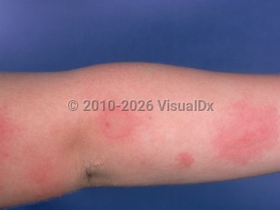
Solar urticaria presents with classic urticarial wheals, erythema, and/or edema limited to sun-exposed areas of the body within minutes of sun exposure. Lesions typically resolve within 1-2 hours after exposure. Regularly sun-exposed areas, such as the face and dorsal hands, may be less sensitive due to the hardening phenomenon. Fixed solar urticaria is a subtype in which lesions occur exclusively in the same localized area of the skin.

Most patients experience accompanying pruritus or a burning sensation, and rarely pain has been described. Systemic symptoms are uncommon but may occur after prolonged exposure of a large body surface area to inciting radiation; symptoms may include headache, nausea, dizziness, wheezing, dyspnea, and syncope. Rarely, severe attacks are associated with anaphylactic shock.

Solar urticaria is caused by immunoglobulin E (IgE)-mediated mast cell degranulation. However, the exact mechanism triggering the degranulation is not known. It is thought to be an IgE-mediated response to a photoinduced allergen. Chlorpromazine, tar, benoxaprofen, and repirinast are known causes of drug-induced solar urticaria.

Solar urticaria typically follows a chronic course. The mean age of onset is 35 years, with less than 4% of patients presenting before age 5. Spontaneous resolution may occur in 15% of patients after 5 years and approximately 25% of patients after 10 years of symptoms. Occasionally, repeat exposure may produce a hardening effect with decreased severity or frequency of symptoms.

Codes

ICD10CM:
L56.3 – Solar urticaria

SNOMEDCT:
10347006 – Solar urticaria

Look For

Subscription Required

Diagnostic Pearls

Subscription Required

Differential Diagnosis & Pitfalls

  • Other forms of urticaria, especially heat-induced urticaria (see physical urticaria). Careful history taking should help differentiate idiopathic from inducible urticaria. The use of a water filter in front of the light source during phototesting helps to rule out heat-induced urticaria.
  • Polymorphous light eruption occurs within hours, not minutes, of sun exposure, and lesions last for days, not minutes or hours, as in solar urticaria. Whereas solar urticaria may occur on the face, polymorphic light eruption usually spares the face.
  • Porphyrias also have lesions on sun-exposed skin. Erythropoietic protoporphyria (EPP) would be clinically most similar to solar urticaria in children. EPP can be differentiated in that it manifests during early childhood; it is typically painful rather than pruritic; petechial, purpuric, hemorrhagic crusted, and/or scarring lesions may be present; and it may have increased protoporphyrin in plasma and red blood cells. Patients with EPP typically have a positive family history, as the disorder is inherited in an autosomal dominant fashion. Porphyria cutanea tarda, also in the differential diagnosis for adults, manifests with blisters and erythema on sun-exposed skin, with scarring and elevated uroporphyrinogen in the urine.
  • Systemic lupus erythematosus should be excluded in all patients with photoinduced skin eruptions. Subacute cutaneous lupus erythematosus spares the midface. Both acute and subacute lupus erythematosus spare the knuckles and may be scaly. Lupus erythematosus tumidus may have urticarial plaques, but lesions last longer than hours.
  • Brachioradial pruritus presents with localized pruritus of the extensor forearm, upper arm, or neck without an associated rash. Sunlight is a common reported trigger. Like solar urticaria, symptoms present commonly in middle-aged females. Burning and stinging may accompany pruritus, and symptoms are often relieved with an ice pack.
  • Photoallergic reactions are type 4 hypersensitivity reactions that present with eczematous dermatitis in sun-exposed areas of the body. Symptoms present 1-2 days after sun exposure, in contrast to solar urticaria. See drug-induced photoallergic reaction.
  • Phototoxic drug reactions occur after ingestion of a photosensitizing agent. Like solar urticaria, the reaction occurs within minutes to hours of sun exposure. However, presentation resembles a sunburn instead of urticaria. Since the reaction is not immune mediated, it will resolve with removal of the offending drug.

Best Tests

Subscription Required

Management Pearls

Subscription Required

Therapy

Subscription Required

Drug Reaction Data

Below is a list of drugs with literature evidence indicating an adverse association with this diagnosis. The list is continually updated through ongoing research and new medication approvals. Click on Citations to sort by number of citations or click on Medication to sort the medications alphabetically.

Subscription Required

References

Subscription Required

Last Reviewed:01/14/2019
Last Updated:12/13/2020
Copyright © 2023 VisualDx®. All rights reserved.
Solar urticaria in Infant/Neonate
A medical illustration showing key findings of Solar urticaria : Burning skin sensation, Edema, Erythema, Sun exposure - recent intense, Blanching patch, Pruritus, Sun-exposed distribution, Urticaria
Clinical image of Solar urticaria - imageId=879551. Click to open in gallery.
Copyright © 2023 VisualDx®. All rights reserved.