Staphylococcal scalded skin syndrome in Child
Alerts and Notices
Important News & Links
Synopsis

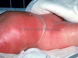
Staphylococcal scalded skin syndrome (SSSS), or Ritter disease, is an acute disease caused by epidermolytic toxins released by strains of Staphylococcus aureus. Any systemic or cutaneous infection with epidermolytic toxin producing S. aureus may induce SSSS. Oftentimes, it is a focal infection of the nasopharynx, conjunctivae, perineum, or umbilicus that produces toxins that lead to diffuse fragile bullae, which are often no longer intact at the time of presentation. Children younger than 6 are believed to have increased susceptibility to SSSS due to decreased renal ability to excrete the toxin. The severity ranges from limited cutaneous involvement to diffuse skin disease and sepsis.
Codes
ICD10CM:
L00 – Staphylococcal scalded skin syndrome
SNOMEDCT:
200946001 – Staphylococcal scalded skin syndrome
L00 – Staphylococcal scalded skin syndrome
SNOMEDCT:
200946001 – Staphylococcal scalded skin syndrome
Look For
Subscription Required
Diagnostic Pearls
Subscription Required
Differential Diagnosis & Pitfalls

To perform a comparison, select diagnoses from the classic differential
Subscription Required
Best Tests
Subscription Required
Management Pearls
Subscription Required
Therapy
Subscription Required
References
Subscription Required
Last Reviewed:06/08/2017
Last Updated:06/14/2017
Last Updated:06/14/2017
Staphylococcal scalded skin syndrome in Child