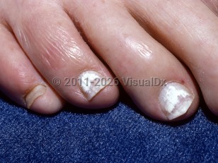
Superficial onychomycosis is a type of fungal nail infection in which the infecting organism invades the upper surface of the nail plate. It usually presents with superficial white patches on the nail plate. Less commonly, it may appear in a transverse striate or invasive pattern. Rarely, it can also have a black color. The scale can often be scraped off with a blade for diagnostic purposes. The most common organisms responsible for the patchy form are Trichophyton rubrum, Trichophyton mentagrophytes, Fusarium, and Acremonium. In the transverse form, Scytalidium, T. rubrum, and Fusarium are often isolated.
Onychomycosis affects up to 14% of the US population. Superficial onychomycosis is a rare subtype that is estimated to account for 1.5%-7% of cases of onychomycosis.
Superficial onychomycosis is usually seen in adults, and it is more common in immunocompromised patients, including those with human immunodeficiency virus (HIV)-infection, those with diabetes, and the elderly. Risk factors for onychomycosis and tinea pedis include hyperhidrosis, wearing occlusive footwear, and walking barefoot through public shower areas, such as in locker rooms.
Superficial white onychomycosis is generally asymptomatic, but may become painful or have a negative effect on quality of life in untreated cases. There is a high association with tinea pedis (athlete's foot), which may be pruritic. Often the initial presentation will be tinea pedis that progresses to onychomycosis over a course of weeks to months.
Pediatric Patient Considerations:
Superficial onychomycosis is rare in children.
Immunocompromised Patient Considerations:
Superficial onychomycosis is associated with an immunocompromised state, particularly HIV-infection.
Superficial onychomycosis - Nail and Distal Digit
Alerts and Notices
Important News & Links
Synopsis

Codes
ICD10CM:
B35.1 – Tinea unguium
SNOMEDCT:
417583002 – Superficial white onychomycosis
B35.1 – Tinea unguium
SNOMEDCT:
417583002 – Superficial white onychomycosis
Look For
Subscription Required
Diagnostic Pearls
Subscription Required
Differential Diagnosis & Pitfalls

To perform a comparison, select diagnoses from the classic differential
Subscription Required
Best Tests
Subscription Required
Management Pearls
Subscription Required
Therapy
Subscription Required
References
Subscription Required
Last Reviewed:08/29/2017
Last Updated:10/03/2017
Last Updated:10/03/2017
Superficial onychomycosis - Nail and Distal Digit