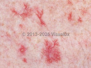
Telangiectasias are permanently visible, dilated, superficial blood vessels, such as a capillary, an arteriole, or a venule. Telangiectasias vary in diameter, up to 1 mm, and range in color from light red to deep purple. The vessels may be linear, branching (arborizing), or spider-like in configuration. Their distribution and associated features help distinguish between different etiologic types.
Telangiectasias may be primary or occur secondary to cutaneous damage, congenital or acquired conditions, or hormonal or metabolic influences. Telangiectasias are also part of the tetrad of findings in poikiloderma, along with atrophy, hyperpigmentation, and hypopigmentation.
The most common types of telangiectasia include facial telangiectasias and those associated with venous hypertension.
Risk factors for facial telangiectasias include aging, chronic photodamage, smoking, female sex, and light skin. Facial telangiectasias may also be a feature of rosacea. Telangiectasias are commonly seen at the nasal alar grooves. Ultraviolet (UV)-induced telangiectasias display a predominantly lateral facial distribution, most commonly involving the preauricular regions and lateral cheeks, whereas those associated with rosacea have a predominantly centrofacial distribution.
Telangiectasias may also be an early sign of venous insufficiency. These are dilated intradermal veins with a diameter less than 1 mm. They may be accompanied by reticular veins (dilated subcutaneous veins with a diameter less than 3 mm) and may accompany varicose veins (tortuous and with a diameter over 3 mm). The precise physiopathology remains unknown, although insufficient perforating veins are found in association with telangiectasias.
Telangiectasia in Adult
Alerts and Notices
Important News & Links
Synopsis

Codes
ICD10CM:
I78.1 – Nevus, non-neoplastic
SNOMEDCT:
1197432002 – Telangiectasia of skin
I78.1 – Nevus, non-neoplastic
SNOMEDCT:
1197432002 – Telangiectasia of skin
Look For
Subscription Required
Diagnostic Pearls
Subscription Required
Differential Diagnosis & Pitfalls

To perform a comparison, select diagnoses from the classic differential
Subscription Required
Best Tests
Subscription Required
Management Pearls
Subscription Required
Therapy
Subscription Required
Drug Reaction Data
Subscription Required
References
Subscription Required
Last Reviewed:04/26/2026
Last Updated:04/29/2026
Last Updated:04/29/2026
 Patient Information for Telangiectasia in Adult
Premium Feature
VisualDx Patient Handouts
Available in the Elite package
- Improve treatment compliance
- Reduce after-hours questions
- Increase patient engagement and satisfaction
- Written in clear, easy-to-understand language. No confusing jargon.
- Available in English and Spanish
- Print out or email directly to your patient
Upgrade Today

Telangiectasia in Adult