David Adelson MD
Christine Ahn MD
Sanah Ali MD
Carl Allen DDS, MSD
Brandon Ayres MD
Howard P. Baden MD
Yevgeniy Balagula MD
Robert Baran MD
Keira Barr MD
Sid Barsky MD
Gregory J. Basura MD, Ph.D
Donald Belsito MD
Jeffrey D. Bernhard MD
Jesse Berry MD
Victor Blanco MD
Benjamin R. Bohaty MD
William Bonnez MD
Mina Botros MD
Jeremy S. Boyd MD
Sarah Brenner MD
Robert A. Briggaman MD
Robert Brodell MD
Roman Bronfenbrener MD
Walter Brooks MD
Daniel Bryan MD
William Buckley MD
Philip Bulterys MD, PhD
Susan Burgin MD
Sonya Burton MD
Sean P. Bush MD, FACEP
Jeffrey Callen MD
Scott Camazine MD
Michael Cardwell
Shelley D. Cathcart MD
Robert Chalmers MD, MRCP, FRCP
Chia-Yu Chu MD, PhD
Flavio Ciferri MD
Maria Rosa Cordisco MD
Noah Craft MD, PhD
John T. Crissey MD
Harold E. Cross MD, PhD
Charles E. Crutchfield III MD
Adriana Cruz MD
Donna Culton MD, PhD
Bart J. Currie MBBS, FRACP, DTM&H
Chicky Dadlani MD
Alexander Dane DO
Roxana Daneshjou MD
C. Ralph Daniel III MD
Thomas Darling MD, PhD
Steven Daveluy MD
William Delaney MD
Shreya Deoghare MD
Alex Derstenfeld
Damian P. DiCostanzo MD
Benedict F. DiGiovanni MD
Ncoza Dlova MD
Erin Doudt
James Earls MD
Libby Edwards MD
Melissa K. Egge MD
Charles N. Ellis MD
Rachel Ellis MD
David Elpern MD
Nancy Esterly MD
Stephen Estes MD
E. Dale Everett MD
Janet Fairley MD
David Feingold MD
Jennifer J. Findeis-Hosey MD
Benjamin Fisher MD
Robert Warne Fitch, MD
Henry Foong MBBS, FRCP
David Foster MD, MPH
Lindy P. Fox MD
Brian D. Foy PhD
Michael Franzblau MD
Vincent Fulginiti MD
Sunir J. Garg MD, FACS
Kevin J. Geary MD
Sharon Glick MD
Lowell Goldsmith MD, MPH
Sethuraman Gomathy MD
Bernardo Gontijo MD, PhD
Ayman Grada MD
Kenneth Greer MD
Kenneth G. Gross MD
Alan Gruber MD
Nathan D. Gundacker MD
Akshya Gupta MD
Vidal Haddad MSC, PhD, MD
Edward Halperin MD, MA
Ronald Hansen MD
John Harvey
Rizwan Hassan MD
Michael Hawke MD
Jason E. Hawkes MD
Peter W. Heald MD
Martha Hickman MD
David G. Hicks MD
Sarah Hocker DO
Ryan J. Hoefen MD, PhD
Li-Yang Hsu MD
Jennifer Huang MD
William W. Huang MD, MPH, FAAD
Eric F. Ingerowski MD
Sanjana Iyengar MD
Alvin H. Jacobs MD
Randy Jacobs MD, FAAD
Saagar Jadeja MD
Shahbaz A. Janjua MD
Joshua J. Jarvis MD
Noman Javed, MD
Beverly A. Johnson MD
Kit Johnson
Zachary John Jones MD
Harsimran Kaur MD
Robert Kalb MD
Katherine Kaproth-Joslin MD, PhD
Philip J. Katzman MD
A. Paul Kelly MD
Henry Kempe MD
Loren Ketai MD
Sidney Klaus MD
Ashwin Kosambia MD
Jessica A. Kozel MD
Carl Krucke
Mario E. Lacouture MD
Joseph Lam MD
Alfred T. Lane MD
Gerald S. Lazarus MD
Edith Lederman MD
Nahyoung Grace Lee MD
Ronald J. Lee MD
Pedro Legua MD, PhD
Robert Levin MD
Bethany Lewis MD
Sue Lewis-Jones FRCP, FRCPCH
Taisheng Li MD
Christine Liang MD
Shari Lipner MD, PhD
Adam Lipworth MD
Jenna Lullo MD
Jason Maguire MD
Mark Malek MD, MPH
Jere Mammino DO
Ricardo Mandojana MD
Lynne Margesson MD
Thomas J. Marrie MD
Maydel Martinez MD
Ralph Massey MD
Patrick McCleskey MD
Lynn McKinley-Grant MD
Karen McKoy MD
Thomas McMeekin MD
Josette McMichael MD
Somchai Meesiri MD
Joseph F. Merola MD
Mary Gail Mercurio MD
Anis Miladi MD
Larry E. Millikan MD
Dan Milner Jr. MD
Zaw Min MD
Nequesha Mohamed MD
Stephanie Montero
Alastair Moore MD
Martha Alejandra Morales-Sánchez MD
Keith Morley MD
Dean Morrell MD
Samuel Moschella MD
Surya N. Mundluru MD
Nicholas Erick Nacca MD
Rehan Naseemuddin MD
Taimor Nawaz MD
Vic Newcomer MD
John Nguyen MD
Matilda Nicholas MD, PhD
Thomas P. Nigra MD
Dylan Norton MD
Steven Oberlender MD, PhD
Maria Teresa Ochoa MD
Edward O'Keefe ND
Daniel C. Oppenheimer MD
Art Papier MD
Lawrence Parish MD
Tanner Parrent MD
Mukesh Patel MD
Lauren Patty-Daskivich MD
David Peng MD, MPH
Robert Penne MD
Nitipong Permpalung MD
Frank Johannes Plate MD, PhD
Miriam Pomeranz MD
Doug Powell MD
Harold S. Rabinovitz MD
Christopher J. Rapuano MD
Sireesha Reddy MD
Angela Restrepo MD, PhD
Cynthia Reyes-Barron MD
Bertrand Richert MD, PhD
J. Martin Rodriguez, MD, FACP
Theodore Rosen MD
Misha Rosenbach MD
Scott Schiffman MD
Robert H. Schosser MD
Glynis A. Scott MD
Carlos Seas MD, MSc
Deniz Seçkin MD
Daniel Sexton MD
Paul K. Shitabata MD
Tor Shwayder MD, FAAP, FAAD
Daniel Siegel, MD
Elaine Siegfried MD
Gene Sienkiewicz MD
Christye Sisson
Jonathan Soh MD
Philip I. Song MD
Mary J. Spencer MD, FAAP
Lawrence B. Stack MD
Sarah Stein MD
William Van Stoecker MD
Frances J. Storrs MD
Erik J. Stratman MD
Lindsay C. Strowd MD
Erika Summers MD
Belinda Tan MD, PhD
Robert Tomsick MD
Hensin Tsao MD, PhD
Richard P. Usatine MD
Jenny Valverde MD
Vishalakshi Viswanath MD
Susan Voci MD
Lisa Wallin ANP, FCCWS
Douglas Walsh MD
Ryan R. Walsh MD
George Watt MD
Erin X. Wei MD
Zachary Werle DO
Clayton E. Wheeler MD
Sally-Ann Whelan MS, NP, CWOCN
Danielle Wilbur MD
Jan Willems MD, PhD
Victoria L. Williams MD
James Henry Willig MD, MPH
Kelly Wilmas MD
Karen Wiss MD
Vivian Wong MD, PhD
Sook-Bin Woo MS, DMD, MMSc
Jamie Woodcock MD
Stephen J. Xenias MD
Nathaniel Yohannes
Lisa Zaba MD
Vijay Zawar MD
Bonnnie Zhang MD
Carolyn Ziemer MD
Barret Zimmerman MD
Jeffrey P. Zwerner MD, PhD
Organizations
American Academy of Dermatology
Am. Journal of Trop. Med & Hygiene
Armed Forces Pest Management Board
Blackwell Publishing
Broward Health Medical Center
Bugwood Network
Centers For Disease Control and Prevention
Centro Internacional de Entrenamiento e Investigaciones Mèdicas (CIDEIM)
Dermatology Associates of Concord
Dermatology Online Journal
East Carolina University (ECU), Division of Dermatology
International Atomic Energy Agency
Massachusetts Medical Society
NYU Department of Dermatology
Oregon Health & Science University (OHSU)
Oxford University Press
Radiological Society of North America
Washington Hospital Center
Wikipedia
World Health Organization
Immunocompetent
Toxoplasmosis is a worldwide zoonosis caused by Toxoplasma gondii, an obligate intracellular protozoan parasite. Toxoplasma gondii exists in 3 morphologic forms: oocyst, tissue cyst (contains bradyzoites), and tachyzoite (the proliferative form). Felines are the definitive host. Mammals (herbivores and carnivores), birds, and reptiles can also be infected. Sexual reproduction takes place only in the feline intestinal tract, resulting in oocysts that are shed in stools.
Human infection may occur from the following:
Ingestion of oocysts in contaminated food or water
Ingestion of tissue cysts contained in undercooked meat (particularly pork, lamb, bear, and deer) or from tissue cysts in transplanted organs (kidney and heart)
Intake of tachyzoites (congenital infection and, rarely, from blood transfusions or laboratory accidents)
On entering the human gastrointestinal (GI) tract, T. gondii infects the gut epithelium and disseminates widely throughout the body, with a predilection for the brain, heart and skeletal muscles, and eyes. The replication of the tachyzoites causes host cell death, and the clinical symptoms and signs depend on the organs involved and the extent of infection and host immunity. Once an effective host immune response has occurred, tachyzoites are killed, while tissue cysts remain and are responsible for the chronic, latent, asymptomatic infection that ensues. This latent infection can be detected serologically. The tissue cysts have a predilection for the central nervous system (CNS), eye, and muscle and serve as a reservoir from which local or disseminated disease can develop if immunosuppression occurs. Reactivation disease usually presents as toxoplasma encephalitis or chorioretinitis.
The incidence of toxoplasmosis increases with increasing age but does not vary significantly by sex. The seroprevalence of antibodies to T. gondii in the United States is about 20%. Seroprevalence rates are much higher in Europe, Africa, and Central and South America.
Acute primary toxoplasmosis in the immunocompetent host most commonly causes an asymptomatic infection (80%-90%). Cervical lymphadenopathy, particularly of the posterior cervical nodes, without other associated signs or symptoms is the most common clinical manifestation. Other lymph node groups may also be involved. Enlarged nodes are usually smooth, firm, mobile, and nontender and do not suppurate. The lymphadenopathy resolves spontaneously over weeks to months. It has been estimated that up to 7% of clinically significant lymphadenopathies are caused by toxoplasmosis.
Very rarely a mononucleosis-like syndrome may occur with headache, fever, sore throat, lymphadenopathy, fatigue, myalgia, arthralgias, malaise, hepatosplenomegaly, macular erythematous rash (during the first week of illness), mild elevations in the serum transaminases, and the presence of atypical lymphocytes on the peripheral blood smear. Some patients may present with only a few of the above features. It is thought that less than 1% of mononucleosis-like syndromes are caused by acute toxoplasmosis. Very rarely, severe disseminated toxoplasmosis may occur in an apparently healthy adult. Depending on the specific organ involved, it may present as encephalitis, myocarditis, hepatitis, polymyositis, or pneumonitis.
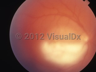
Ocular toxoplasmosis can result from congenital infection or from reactivation of latent postnatally acquired infection and rarely develops during acute infection. Toxoplasma gondii is the most common cause of chorioretinitis in immunocompetent adults. The prevalence of ocular toxoplasmosis is unknown but has been estimated at 2% of seropositive patients. Relapse, even after therapy, particularly in the eye initially involved, is the rule. Patients may complain of blurry vision, scotoma, photophobia, redness, and excessive lacrimation. Atypical findings include eye pain, prominent retinal hemorrhage, and lack of retinal scarring on ophthalmologic examination. They warrant polymerase chain reaction (PCR) testing of the vitreous or aqueous fluid for toxoplasma DNA to confirm the diagnosis.
Pregnant Patient Considerations: Acute T. gondii infection in pregnant individuals is usually asymptomatic. The most frequent clinical manifestation is regional lymphadenopathy. Acute maternal infection can result in transmission to the fetus. However, the risk of fetal transmission is not increased if maternal infection is symptomatic. With rare exceptions (infection within 3 months of conception or maternal immunosuppressed state), women previously infected with T. gondii, and therefore seropositive for anti-toxoplasma antibodies prior to pregnancy, are protected from transmitting the infection to their fetuses. HIV-infected women with latent toxoplasmosis may have reactivation of latent infection, which can result in fetal transmission. The incidence of fetal transmission is highest during second- and third-trimester maternal infection. Though less frequent, fetal transmission during first-trimester maternal infection more commonly results in severe illness in the newborn. See congenital toxoplasmosis.
Immunocompromised Patient Considerations: Seronegative organ transplant recipients from a T. gondii-seropositive donor, seropositive hematopoietic stem cell transplant recipients, patients with AIDS, and those receiving corticosteroids are at risk for disease caused by reactivation of chronic latent T. gondii infection. Primary toxoplasmosis is very rare but can cause disseminated disease, which is frequently fatal. The most common presentation of reactivation of latent infection is toxoplasmic encephalitis and much less frequently chorioretinitis; other organ involvement (pneumonitis, myocarditis) occurs rarely. Manifestations of toxoplasmic encephalitis include focal neurological deficits (hemiparesis, speech abnormalities, altered mental status, movement disorders), seizures, headache, and fever.
Acute cytomegalovirus (CMV) infection – Check for CMV IgM positivity in the serum and the presence of CMV DNA in the blood by PCR testing.
Acute HIV infection – Elicit exposure risk factors. Standard HIV testing (ELISA) may be negative at first but will become positive within a few weeks. Check an HIV viral load, which is always positive in this setting.
Lymphadenopathy without other associated symptoms – Think about lymphomalymphoma, and sample a lymph node (fine needle aspiration or excisional biopsy).
Cat-scratch disease – Bartonella henselae (and the putative agents Afipia felis and Bartonella clarridgeiae); serology can establish the diagnosis.
Ocular Toxoplasmosis:
Ocular toxoplasmosis may resemble the chorioretinitis of tuberculosis; check purified protein derivative (PPD) and chest X-ray.
In patients with atypical lesions or a poor response to anti-toxoplasma therapy, consider toxoplasma DNA detection by PCR in the vitreous or aqueous fluid.
Immunocompromised Patient Differential Diagnosis:
Primary CNS non-Hodgkin lymphoma of the brain is by far the most common entity in the differential diagnosis of toxoplasmic encephalitis. Patients virtually always have CD4 cell counts <50/mm3. Serology for toxoplasmosis is usually negative. CSF EBV DNA PCR detection is usually positive. Thallium-201 single proton emission CT (SPECT) scanning shows increased metabolic activity. A definitive diagnosis may require brain biopsy.