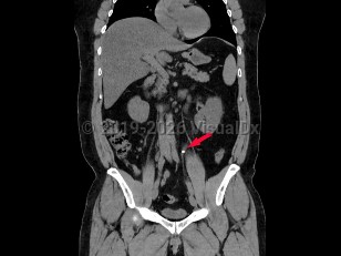
Ureteral calculi are stones that have formed in the kidney and passed from the renal collecting system into the ureter. For a more detailed discussion on the demographics, etiology and pathophysiology, and clinical associations of nephrolithiasis, please see renal calculus.
While many stones located within the renal pelvis are discovered incidentally on radiographic or sonographic imaging, most stones that have passed into the ureter and are of significant size cause renal colic. Renal colic is sudden-onset flank pain radiating to the groin, accompanied by nausea and vomiting. This colicky pain typically waxes over the course of 15-30 minutes and becomes steady, unrelenting, and unbearable. Patients may experience worsening paroxysms of pain lasting 20-60 minutes as the stone courses downward through the ureter and as the ureter spasms.
If the stone's descent is arrested at the ureterovesical junction, patients may experience urinary frequency, dysuria, and urgency and are predisposed to the development of urinary tract infections both from the stone forming as a nidus for bacterial growth and from the mechanical urothelial trauma caused by the stone's movement. Most individuals with nephrolithiasis will also develop hematuria, particularly when passing a stone.
Pain from nephrolithiasis is thought to primarily be the result of renal capsular distention and varies depending on the location of the stone and the degree of obstruction caused by the stone. Stones that occlude the upper ureter or ureteropelvic junction invariably cause significant flank pain that is accompanied by severe costovertebral angle tenderness to palpation. As the innervation of the testicle is shared with the kidney, patients often describe radiation to the testicles or labia. When stones pass into the bladder, patients usually experience swift resolution of their pain.
Ureteral calculus
Alerts and Notices
Important News & Links
Synopsis
Codes
ICD10CM:
N20.1 – Calculus of ureter
SNOMEDCT:
31054009 – Ureteric stone
N20.1 – Calculus of ureter
SNOMEDCT:
31054009 – Ureteric stone
Look For
Subscription Required
Diagnostic Pearls
Subscription Required
Differential Diagnosis & Pitfalls

To perform a comparison, select diagnoses from the classic differential
Subscription Required
Best Tests
Subscription Required
Management Pearls
Subscription Required
Therapy
Subscription Required
Drug Reaction Data
Subscription Required
References
Subscription Required
Last Reviewed:08/12/2018
Last Updated:08/12/2018
Last Updated:08/12/2018
Ureteral calculus