Mastocytosis is a term broadly referring to tissue mast cell hyperplasia. According to the World Health Organization (WHO) classification, cutaneous and systemic forms of mastocytosis are distinguished. Mastocytosis most commonly manifests as cutaneous disease in childhood, which is further subdivided into urticaria pigmentosa (UP, also known as maculopapular cutaneous mastocytosis), localized mastocytoma of skin, and diffuse cutaneous mastocytosis. Cutaneous mastocytosis in childhood has an excellent prognosis, with many cases remitting during puberty. Severe presentations may be persistent into adulthood (see mastocytosis in adults).
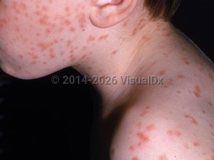
UP is the most common form of cutaneous mastocytosis in children. UP presents with few to many persistent, pink and brown macules and papules that urticate or wheal with thermal, mechanical, or chemical stimuli. Up to 25% of pediatric UP patients develop gastrointestinal symptoms. Fever, night sweats, malaise, epigastric tenderness, weight loss, flushing, diarrhea, syncope, bronchospasm, and problems with cognition signal possible systemic involvement.
Somatic c-kit mutations and other mast cell immunophenotypic alterations have been implicated in the pathophysiology of mastocytosis.
When triggered, mast cells release mediators such as histamine prostaglandins, eicosanoids, heparin, leukotrienes, cytokines, and proteases, causing patients with UP to become symptomatic. Triggers can include heat or temperature changes, trauma to skin lesions, insect stings, psychological stress, anxiety, and certain drugs such as NSAIDs, anesthetics, opioids, muscle relaxants, antibiotics, and iodinated radiocontrast agents.
UP can present at any time, but it commonly presents within the first weeks to months of life and can be expected to spontaneously involute by adolescence. There is no sex or race predilection. Partial to complete regression occurs in most children, as opposed to adults, in whom UP tends to persist.
Urticaria pigmentosa in Infant/Neonate
Alerts and Notices
Important News & Links
Synopsis

Codes
ICD10CM:
Q82.2 – Mastocytosis
SNOMEDCT:
34739009 – Cutaneous mastocytosis, adult form
Q82.2 – Mastocytosis
SNOMEDCT:
34739009 – Cutaneous mastocytosis, adult form
Look For
Subscription Required
Diagnostic Pearls
Subscription Required
Differential Diagnosis & Pitfalls

To perform a comparison, select diagnoses from the classic differential
Subscription Required
Best Tests
Subscription Required
Management Pearls
Subscription Required
Therapy
Subscription Required
Drug Reaction Data
Subscription Required
References
Subscription Required
Last Reviewed:03/23/2020
Last Updated:03/26/2020
Last Updated:03/26/2020
Urticaria pigmentosa in Infant/Neonate