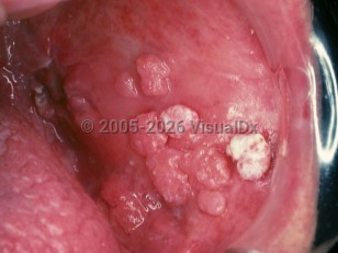
Oral verrucous carcinoma (also known as Ackerman tumor) is a rare, indolent variant of oral squamous cell carcinoma (SCC), representing approximately 5% of SCCs and 3%-4% of all oral carcinomas. Characteristically, oral verrucous carcinomas present as white or pink, painless, velvety lesions or warty exophytic tumors in the oral cavity (buccal mucosa and gingiva), oropharynx, and/or larynx.
Noninvasive verrucous mucosal lesions develop into proliferative verrucous leukoplakia, which can then progress to oral verrucous carcinoma or typical oral SCC.
Verrucous carcinomas rarely metastasize and have favorable prognoses. However, verrucous carcinomas can grow and destroy adjacent tissue, cartilage, and bone if left untreated. Patients usually report being aware of the lesion or tumor for years prior to seeking evaluation by a medical professional.
Men over the age of 55 most commonly develop oral verrucous carcinoma. Women may also develop verrucous carcinoma, but with a different presentation. In a 2001 study, women were reported to have a higher proportion of oral cavity tumors whereas men have higher proportion of laryngeal tumors.
Although most studies report an increased incidence of mucosal verrucous carcinoma among tobacco users (including dry and inhaled tobacco products), 16%-51% of oral verrucous carcinomas have been reported in patients without a history of tobacco use. When tobacco use is present and verrucous carcinoma is suspected, the mandibular vestibule (placement site for dry tobacco), buccal mucosa, hard palate, oropharynx, and larynx should be carefully inspected.
Human papillomavirus (HPV) has not been proven to be associated with oral verrucous carcinoma despite HPV having a proven role in many other verrucous conditions and verrucous carcinomas in other locations.
Verrucous carcinoma - Oral Mucosal Lesion
See also in: OverviewAlerts and Notices
Important News & Links
Synopsis

Codes
ICD10CM:
C44.320 – Squamous cell carcinoma of skin of unspecified parts of face
D04.9 – Carcinoma in situ of skin, unspecified
SNOMEDCT:
89906000 – Verrucous Carcinoma
C44.320 – Squamous cell carcinoma of skin of unspecified parts of face
D04.9 – Carcinoma in situ of skin, unspecified
SNOMEDCT:
89906000 – Verrucous Carcinoma
Look For
Subscription Required
Diagnostic Pearls
Subscription Required
Differential Diagnosis & Pitfalls

To perform a comparison, select diagnoses from the classic differential
Subscription Required
Best Tests
Subscription Required
Management Pearls
Subscription Required
Therapy
Subscription Required
References
Subscription Required
Last Reviewed:01/17/2018
Last Updated:01/30/2018
Last Updated:01/30/2018
Verrucous carcinoma - Oral Mucosal Lesion
See also in: Overview