Warty dyskeratoma, or follicular dyskeratoma, is a relatively uncommon, benign, follicular adnexal neoplasm that usually appears between the fifth and seventh decades of life and is most prevalent in White men. The pathogenesis remains uncertain, but ultraviolet (UV) light, abnormal adhesion of keratinocytes, autoimmunity, and smoking may play a role. Architectural features resembling a wart-like growth have led to the suggestion of a viral infection to be the cause. However, human papillomavirus (HPV) infection has not been detected in these lesions.
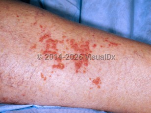
Lesions usually present as a slow-growing, solitary, well-demarcated, skin-colored-to-red-brown umbilicated papule or nodule with a crusted, keratotic center. Most are asymptomatic; rarely, patients complain of pruritus or burning, and bleeding, crusting, or drainage may occur.
Warty dyskeratomas are most commonly located on sun-exposed areas of the body (scalp, face, and neck). They have been documented to coexist with other skin lesions, including actinic keratosis, squamous cell carcinoma (SCC), basal cell carcinoma (BCC), and verruciform xanthoma. Multiple lesions, lesions involving the oral and vulvar mucosa, and subungual involvement have occasionally been reported. Eruptive warty dyskeratomas have been reported to occur in 2 patients with end-stage renal disease.
Warty dyskeratomas are benign. Malignant transformation has not been reported.
Warty dyskeratoma
See also in: Hair and ScalpAlerts and Notices
Important News & Links
Synopsis

Codes
ICD10CM:
L85.8 – Other specified epidermal thickening
SNOMEDCT:
254676008 – Warty dyskeratoma
L85.8 – Other specified epidermal thickening
SNOMEDCT:
254676008 – Warty dyskeratoma
Look For
Subscription Required
Diagnostic Pearls
Subscription Required
Differential Diagnosis & Pitfalls

To perform a comparison, select diagnoses from the classic differential
Subscription Required
Best Tests
Subscription Required
Management Pearls
Subscription Required
Therapy
Subscription Required
References
Subscription Required
Last Reviewed:02/02/2022
Last Updated:02/15/2022
Last Updated:02/15/2022
Warty dyskeratoma
See also in: Hair and Scalp