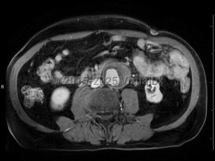
Hemodynamically unstable patient:
- Two large bore intravenous (IV) catheters
- Blood pressure control
- Pain control
- Computed tomography angiography (CTA) imaging is preferred. Testing for renal function is not necessary if there is a high index of suspicion for abdominal aortic aneurysm (AAA); there is a weak association between contrast and acute kidney injury (AKI), and the risks outweigh the benefits.
- Consultation with vascular surgery does not need to be delayed by imaging confirmation in the setting of an unstable patient with a high index of suspicion.
- Bedside ultrasound may show a positive focused assessment with sonography in trauma (FAST) examination in patients with a ruptured AAA, although it may be negative if the bleed is retroperitoneal. Ultrasound is not sensitive for rupture.
- The aorta diameter is measured from outer wall to outer wall. A common pitfall is the misinterpretation of thrombus with the outer wall of the aorta.
AAA is defined as a focal, full-thickness dilation of the abdominal aorta that is 50% or more of the regular diameter, or 3 cm or larger. The abdominal aorta is retroperitoneal and lies between the diaphragm and aortic bifurcation, with about 80% of aneurysms arising infrarenally. They are most commonly degenerative in patients with atherosclerosis; 5%-10% are inflammatory and are more often symptomatic.
AAAs are most commonly diagnosed in men with a smoking history, but other risk factors for development are advanced age, other aneurysm, atherosclerotic disease, hypertension, hypercholesterolemia, chronic obstructive pulmonary disease (COPD), a family history of AAA, and White race. In women, 3 cm or larger is aneurysmal, but aortic size index (ASI, diameter in cm / body surface area [BSA]) is a better predictor of clinical events than diameter alone.
General size classifications:
- Small: < 4 cm
- Medium: 4-5.5 cm
- Large: > 5.5 cm
- Very large: ≥ 6 cm
Identifying an AAA is essential, as mortality associated with rupture is up to 80% and is as high as 50%-70% in patients who make it to the hospital after a rupture. Current guidelines recommend screening for AAA in men aged 65-75 years who have ever smoked and in men or women who have a first-degree relative with an AAA.
Related topic: cystic medial necrosis


