Contents

SynopsisCodesLook ForDiagnostic PearlsDifferential Diagnosis & PitfallsBest TestsManagement PearlsTherapyReferences

Information for Patients

View all Images (6)

Atopic dermatitis - Cellulitis DDx
See also in: Overview,External and Internal Eye
Other Resources UpToDate PubMed

Atopic dermatitis - Cellulitis DDx

See also in: Overview,External and Internal Eye
Contributors: Azeen Sadeghian MD, Ryan Fan BA, Jeffrey M. Cohen MD, Susan Burgin MD
Other Resources UpToDate PubMed

Synopsis

Atopic dermatitis (eczema) is a chronic, relapsing, pruritic condition that is often associated with allergic rhinitis and/or asthma. Infants and children are most often affected, with 85% of cases appearing in the first year of life and 95% of cases appearing by 5 years. However, in fewer individuals the condition may persist into, or even arise in, adulthood. Less than 1% of adults are affected by atopic dermatitis. With increased understanding of immunosenescence, atopic dermatitis is increasingly being recognized in the older adult population.

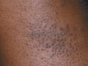
In infants, the disease involves primarily the face, scalp, and torso. In children and adults, the disease usually involves chiefly the flexural aspects of extremities, but it may be more generalized. Follicular patterns of atopic dermatitis are more common in persons of color. A lichen planus-like appearance has also been reported in persons of color, due to lichenification and the difficulties in perceiving erythema in darker skin colors.

Atopic dermatitis may be categorized as follows:
  • Acute – erythema, vesicles, bullae, weeping, crusting
  • Subacute – scaly plaques, papules, round erosions, crusts
  • Chronic eczema – lichenification, scaling, hyper- and hypopigmentation
The cause of atopic dermatitis is unknown. Genetic and environmental predisposing factors exist. Multiple loci have been associated with atopic dermatitis. A family history of atopic dermatitis is common.

Intense pruritus (itching) is a hallmark of atopic dermatitis. Scratching leads to lichenification (skin thickening from chronic trauma). Impaired barrier function leads to increased water loss and cutaneous infections. Patients with atopic dermatitis are prone to impetiginization with Staphylococcus aureus. Secondary infections with herpes simplex virus (eczema herpeticum), Coxsackie viruses (eczema coxsackium), or vaccinia virus (eczema vaccinatum) may transpire.

Patients with atopic dermatitis have difficulties in retaining skin moisture and suffer from xerosis (dry skin). Environmental triggers, such as heat, humidity, detergents / soaps, abrasive clothing, chemicals, smoke, and even stress, tend to aggravate the condition. Latex allergy and nickel allergy occur more often in persons with atopic dermatitis. Additionally, patients with atopic dermatitis have been found to be more likely to have positive patch test results to products commonly found in topical treatments, including cocamidopropyl betaine, wool alcohol / lanolin, and tixocortol pivalate. Allergy to eggs, cow's milk, or peanuts is common. There may be a relationship between atopic dermatitis and the development of aspirin-related respiratory disease.

Atopic dermatitis can be differentiated from cellulitis on the basis of itching, scaling, and distribution. Atopic dermatitis is often observed bilaterally and in multiple body locations.

Codes

ICD10CM:
L20.9 – Atopic dermatitis, unspecified

SNOMEDCT:
24079001 – Atopic dermatitis

Look For

Subscription Required

Diagnostic Pearls

Subscription Required

Differential Diagnosis & Pitfalls

To perform a comparison, select diagnoses from the classic differential

Subscription Required

Best Tests

Subscription Required

Management Pearls

Subscription Required

Therapy

Subscription Required

References

Subscription Required

Last Reviewed:11/20/2023
Last Updated:11/21/2023
Copyright © 2024 VisualDx®. All rights reserved.
Patient Information for Atopic dermatitis - Cellulitis DDx
Print E-Mail Images (6)
Contributors: Medical staff writer
Premium Feature
VisualDx Patient Handouts
Available in the Elite package
  • Improve treatment compliance
  • Reduce after-hours questions
  • Increase patient engagement and satisfaction
  • Written in clear, easy-to-understand language. No confusing jargon.
  • Available in English and Spanish
  • Print out or email directly to your patient
Copyright © 2024 VisualDx®. All rights reserved.
Atopic dermatitis - Cellulitis DDx
See also in: Overview,External and Internal Eye
A medical illustration showing key findings of Atopic dermatitis : Bilateral distribution, Dry skin, Erythema, Excoriated skin lesion, Flexural distribution, Lichenified plaque, Ocular pruritus, Pruritus
Clinical image of Atopic dermatitis - imageId=214627. Click to open in gallery.  caption: 'A close-up of follicular papules with scale.'
A close-up of follicular papules with scale.
Copyright © 2024 VisualDx®. All rights reserved.