Hereditary hemochromatosis (HH) is a genetic disorder of iron overload. In the classic form, a C282Y mutation in the HFE gene inappropriately increases iron absorption from the duodenum. Other forms of HH involve mutations in various molecules that regulate iron homeostasis. While usually autosomal recessive, an autosomal dominant form is caused by a mutation in ferroportin.
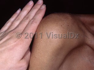
In all forms, excess iron deposits in the joints and in organs such as the liver, pancreas, heart, and skin, causing damage. Historically, the disease is known by the classic triad of hyperpigmentation, cirrhosis, and diabetes mellitus ("bronze diabetes"). Iron deposits also lead to arthropathy (arthritis), edema, hypopituitarism, hypogonadism, hair loss, and cardiac arrhythmias, and advanced liver fibrosis or liver cancer. Individuals are also at increased risk of Vibrio vulnificus and other infections. Patients initially present between the ages of 40 and 60 with nonspecific symptoms such as fatigue, abdominal pain, joint pain, and loss of libido. Hyperpigmentation is eventually seen in about 70% of cases.
There are a few theories regarding the possible mechanism for hyperpigmentation. Deposition of iron in the skin may cause an increase in melanin contained in giant melanosomes. Iron excess may stimulate adrenocorticotropic hormone (ACTH) or melanocyte-stimulating hormone (MSH). Hyperpigmentation may also be a direct manifestation of hemosiderin deposition in the skin.
HFE-associated hemochromatosis most commonly affects White populations, with prevalence in Europe of 1:400. In North America, the prevalence among White people is approximately 1:227. In contrast, non-HFE HH is found worldwide. In southern Europe and Asia, a larger proportion of HH involves non-HFE mutations. It is associated with HLA-A3. Men and women are equally affected, but there are variable clinical manifestations between men and women. Symptoms are more common and often more severe in men, and there is increased mortality in men.
Pediatric patient considerations: Juvenile hemochromatosis (hemochromatosis type 2) is caused by a mutation in the TFR2 gene on chromosome 7. This form is rapidly progressive. Hypogonadism is the typical presenting feature. Cardiac involvement is frequent, and most fatalities are due to heart failure.
Neonatal forms cause liver failure within the first 30 days of life. The condition is likely caused by genetic factors as well as various in utero insults. It presents with jaundice.
Hemochromatosis
Alerts and Notices
Important News & Links
Synopsis

Codes
ICD10CM:
E83.110 – Hereditary hemochromatosis
SNOMEDCT:
399187006 – Hemochromatosis
E83.110 – Hereditary hemochromatosis
SNOMEDCT:
399187006 – Hemochromatosis
Look For
Subscription Required
Diagnostic Pearls
Subscription Required
Differential Diagnosis & Pitfalls

To perform a comparison, select diagnoses from the classic differential
Subscription Required
Best Tests
Subscription Required
Management Pearls
Subscription Required
Therapy
Subscription Required
References
Subscription Required
Last Updated:04/11/2024
Hemochromatosis