Mucous membrane pemphigoid (MMP), also known as cicatricial pemphigoid, is a heterogeneous group of chronic autoimmune blistering diseases caused by autoimmunity to various components of the basement membrane. To date, autoantibodies to over 10 different antigens in the basement membrane zone have been identified: bullous pemphigoid antigen 1 (BP230), bullous pemphigoid antigen 2 (BP180, type XVII collagen), laminin-5 (laminin-332), laminin-6 (laminin-3), type VII collagen, integrin beta 4 subunit of α6β4 integrin, 45-kD unknown epithelial protein, 168-kD unknown epithelial protein, 120-kD unknown epithelial protein, and uncein. Patients with autoantibodies to β4 integrin present predominantly with ocular disease.
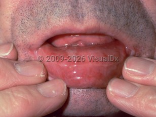
MMP affects the mucous membranes and, less commonly, the skin. The mouth is involved most often, followed by the conjunctiva. MMP causes painful ulcers and erosions in the oral cavity. Because oral hygiene is difficult to perform, gingival lesions are exacerbated by plaque build-up. Patients will often report pain and bleeding on tooth brushing. Patients also avoid hard and spicy foods and may lose weight because of reduced food intake. While cicatrization is common in the larynx, eye, and skin, it is uncommon in the oral mucosa. Oral MMP is almost twice as common in females as it is in males, and it is seen most frequently in older individuals.
If MMP affects the eye, there may be corneal inflammation and scarring, conjunctiva inflammation, trichiasis, ectropion, symblepharon, ankyloblepharon, and blindness. Skin, nasal, anogenital, laryngeal, pharyngeal, and esophageal mucosal surfaces can also be affected, leading to epistaxis, perianal erythema and scarring, phimosis or vaginal scarring, and hoarseness or dysphagia, respectively. Scarring is the endpoint for all sites of involvement except the oral mucosa. Cutaneous disease, when present, most frequently accompanies mucous membrane disease. Occasionally, cutaneous blistering and scarring dominate the clinical picture (so-called Brunsting-Perry variant).
Lesions develop over weeks to months.
In a 2022 study, malignancies, especially solid organ tumors, were reported in up to 13.8% of patients. These include lung carcinoma, prostate cancer, penile cancer, breast cancer (female or male), endometrial cancer, vulvar carcinoma, and non-Hodgkin lymphoma. The malignancy rate was higher when autoantibodies against laminin-332 were found.
Mucous membrane pemphigoid - Oral Mucosal Lesion
See also in: Overview,AnogenitalAlerts and Notices
Important News & Links
Synopsis

Codes
ICD10CM:
L12.1 – Cicatricial pemphigoid
SNOMEDCT:
34250006 – Cicatricial pemphigoid
L12.1 – Cicatricial pemphigoid
SNOMEDCT:
34250006 – Cicatricial pemphigoid
Look For
Subscription Required
Diagnostic Pearls
Subscription Required
Differential Diagnosis & Pitfalls

To perform a comparison, select diagnoses from the classic differential
Subscription Required
Best Tests
Subscription Required
Management Pearls
Subscription Required
Therapy
Subscription Required
Drug Reaction Data
Subscription Required
References
Subscription Required
Last Reviewed:08/09/2022
Last Updated:08/10/2022
Last Updated:08/10/2022
Mucous membrane pemphigoid - Oral Mucosal Lesion
See also in: Overview,Anogenital