Contents

SynopsisCodesLook ForDiagnostic PearlsDifferential Diagnosis & PitfallsBest TestsManagement PearlsTherapyDrug Reaction DataReferences

View all Images (13)

Multifocal or diffuse mucosal pigmentation - Oral Mucosal Lesion
Other Resources UpToDate PubMed

Multifocal or diffuse mucosal pigmentation - Oral Mucosal Lesion

Contributors: Susan Burgin MD, Carl Allen DDS, MSD, Sook-Bin Woo MS, DMD, MMSc
Other Resources UpToDate PubMed

Synopsis

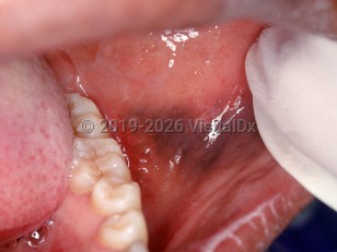
Mucosal pigmentation is a discoloration of the mucosa that may take the form of multifocal macules or diffuse pigmentation caused by either exogenous or endogenous pigmented substances. The most common multifocal or diffuse pigmented state in the oral cavity is physiologic pigmentation that occurs more frequently in individuals with darker skin phototypes.

Mucosal pigmentation caused by exogenous substances can be divided into two categories depending on the pigment: heavy metal pigmentation of systemic origin and medication-induced pigmentation. Heavy metal pigmentation may be caused by lead, arsenic, bismuth, or lead exposure from contaminated opium and often presents as a linear discoloration along the gingival margin (the "lead line" [Burton's line]). Even the bismuth present in Pepto-Bismol can cause a hyperpigmentation, although this is generally localized contact pigmentation on the dorsum of the tongue. It is believed that heavy metal ions excreted through the gingival crevicular fluid react with sulfites produced by gingival and periodontal bacteria, resulting in the deposition of heavy metal sulfides that are often black.

The medications that can cause drug-induced oral pigmentation include minocycline, antimalarials, clofazimine, and oral contraceptives. The drug or drug metabolites are pigmented substances that can be identified lying free or chelated to iron or melanin within the hard and/or soft tissues. Other drugs that supposedly cause oral pigmentation are likely related to a postinflammatory hypermelanosis from a lichenoid interface drug eruption or from direct damage to the mucosa (such as chemotherapeutic agents). Although tetracycline can cause intrinsic staining of the teeth and bone, it does not generally cause mucosal pigmentation.

Mucosal pigmentation caused by endogenous pigment is usually melanotic or hemosiderotic in origin, the latter from the breakdown of blood products. Melanotic pigmentation is seen in Peutz-Jeghers syndrome, McCune-Albright syndrome, hypoadrenal states (such as Addison disease), neurofibromatosis, LEOPARD syndrome, Laugier-Hunziker syndrome, and Carney complex, to name a few.

Codes

ICD10CM:
K13.79 – Other lesions of oral mucosa

SNOMEDCT:
249405005 – Oral pigmentation

Look For

Subscription Required

Diagnostic Pearls

Subscription Required

Differential Diagnosis & Pitfalls

Causes of diffuse oral pigmentation:
  • Physiologic pigmentation – Diffuse and bilateral, childhood onset.
  • Peutz-Jeghers syndrome (periorificial lentiginosis) – Multiple diffuse and bilateral macules on the lips, oral / intranasal / conjunctival / rectal mucosa; childhood onset.
  • Cronkhite-Canada syndrome – Nonfamilial gastrointestinal polyposis syndrome with associated cutaneous hyperpigmented macules, onychodystrophy, and alopecia.
  • Laugier-Hunziker syndrome – Rare, benign condition of macular hyperpigmentation of mucocutaneous surfaces and nails.
  • McCune-Albright syndrome – Polyostotic fibrous dysplasia, café-au-lait spots, and gonadotropin independent precocious puberty.
  • Carney complex – Lentigines of the lips and intraoral pigmentation may be present.
  • Primary hypoadrenalism (Addison disease) – Diffuse blue-black streaks or patches on oral mucosa and tongue in addition to hyperpigmentation, particularly in sun-exposed areas and flexural creases; also with fatigue, hypotension, electrolyte abnormalities.
  • Other endocrine disorders: hyperthyroidism, Cushing disease
  • Drug-induced oral pigmentation including from minocycline, antimalarials, clofazimine, and oral contraceptives
  • Elevated heavy metal levels (lead, bismuth, silver, mercury, gold, arsenic) – Blue-black discoloration along the gingival margin.
  • Post-inflammatory pigmentation – Hyperpigmented lesions at sites of previous inflammatory or traumatic lesions or adjacent to active lesions (eg, lichen planus, pemphigus, pemphigoid).
  • Smoker's melanosis – Brown-black lesions on the anterior labial and buccal mucosa in smokers.
Purple macules may mimic brown pigmented macules:
  • Scurvy – Ecchymosis, sometimes dusky purple discoloration of the gingiva with intraoral swelling and hemorrhage.
  • Kaposi sarcoma – Bilateral brown to purple plaques / nodules in human immunodeficiency virus (HIV)-positive individuals; ulcerative and necrotic in advanced stages.
Differential diagnosis for focal pigmentation:
  • Melanotic macule – Usually less than 1 cm, well demarcated, light or dark brown; more common in women and young adults. Though benign, should consider biopsy to rule out melanoma.
  • Pigmented nevi – Brown or blue macule or papule commonly on the palate. Though benign, should consider biopsy to rule out melanoma.
  • Melanoma – Brown or black patch with irregular borders, rapidly growing and more ulcerated as it advances; commonly on the palate, followed by the gingiva.
  • Hemangioma – Red-bluish / purple lesion, flat or slightly raised, blanches with pressure, most commonly on tongue.
  • Varix – Bluish / purple, irregular elevation, blanches with pressure, commonly on ventral tongue; if it does not blanch with pressure, it may contain a thrombus.
  • Hemorrhage – Nonblanching macule, papule, or petechiae; may be ecchymotic.

Best Tests

Subscription Required

Management Pearls

Subscription Required

Therapy

Subscription Required

Drug Reaction Data

Below is a list of drugs with literature evidence indicating an adverse association with this diagnosis. The list is continually updated through ongoing research and new medication approvals. Click on Citations to sort by number of citations or click on Medication to sort the medications alphabetically.

Subscription Required

References

Subscription Required

Last Reviewed:05/21/2018
Last Updated:05/02/2019
Copyright © 2023 VisualDx®. All rights reserved.
Multifocal or diffuse mucosal pigmentation - Oral Mucosal Lesion
A medical illustration showing key findings of Multifocal or diffuse mucosal pigmentation
Clinical image of Multifocal or diffuse mucosal pigmentation - imageId=2556932. Click to open in gallery.  caption: 'A dark brown patch on the buccal mucosa.'
A dark brown patch on the buccal mucosa.
Copyright © 2023 VisualDx®. All rights reserved.