Basal cell carcinoma (BCC) is the most common cancer in humans and the most common cancer of the skin. Two million Americans are diagnosed with BCC every year. It is a neoplasm of basal keratinocytes that is found more frequently in men than women. Rates of BCC have been increasing over the last several decades, particularly in young women. BCCs can be seen at almost any age. Nonetheless, the malignancy has greater incidence in older individuals, with a median age at diagnosis of 68 years.
The greatest risk factor contributing to the development of BCCs is sun exposure, and people with light skin phototypes are at higher risk. Intermittent sun exposure is more closely associated with the development of BCCs than cumulative ultraviolet exposure.
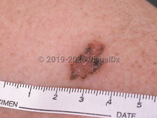
There are many subtypes of BCC, including nodular, superficial, infundibulocystic, fibroepithelial, morpheaform (sclerosing, desmoplastic), infiltrative, micronodular, and basosquamous. Accumulation of melanin and melanophages in the BCC tumor nodules gives rise to clinically pigmented BCCs. Pigmented BCCs account for approximately 6%-7% of nodular, superficial, and micronodular subtypes. They can occur in any location but are most commonly found on the head and neck. They comprise more than 50% of all BCCs in individuals with intermediate and darker skin colors. Pigmented BCCs are observed twice as frequently in Hispanic patients as compared to White patients.
Other risk factors for BCCs include environmental exposure (ie, ionizing radiation, indoor tanning, chemicals such as arsenic, psoralen plus UVA, and coal tar), phenotype (freckling, red hair, light skin that always burns and never tans), immunosuppression such as organ transplantation (which results in a 5-10 times higher risk of BCCs than the general population), and various genetic syndromes including xeroderma pigmentosum, oculocutaneous albinism, Muir-Torre syndrome, basal cell nevus syndrome (Gorlin syndrome), Rombo syndrome, and Bazex-Dupré-Christol syndrome. The gene most frequently altered in BCCs is the PTCH1 gene, followed by the TP53 gene.
Although BCCs are almost never fatal, local tissue destruction and disfiguration do occur. The metastasis rate is approximately 1 in 35 000. Metastasis is rare and typically occurs through perineural spread, lymph node metastasis, and then lung / bone metastasis.
Pigmented basal cell carcinoma
Alerts and Notices
Important News & Links
Synopsis

Codes
ICD10CM:
C44.91 – Basal cell carcinoma of skin, unspecified
SNOMEDCT:
403909004 – Pigmented basal cell carcinoma
C44.91 – Basal cell carcinoma of skin, unspecified
SNOMEDCT:
403909004 – Pigmented basal cell carcinoma
Look For
Subscription Required
Diagnostic Pearls
Subscription Required
Differential Diagnosis & Pitfalls

To perform a comparison, select diagnoses from the classic differential
Subscription Required
Best Tests
Subscription Required
Management Pearls
Subscription Required
Therapy
Subscription Required
References
Subscription Required
Last Reviewed:08/11/2021
Last Updated:01/29/2023
Last Updated:01/29/2023
 Patient Information for Pigmented basal cell carcinoma
Premium Feature
VisualDx Patient Handouts
Available in the Elite package
- Improve treatment compliance
- Reduce after-hours questions
- Increase patient engagement and satisfaction
- Written in clear, easy-to-understand language. No confusing jargon.
- Available in English and Spanish
- Print out or email directly to your patient
Upgrade Today

Pigmented basal cell carcinoma