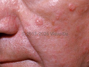
Sebaceous adenomas are benign neoplasms that derive from sebaceous glands. They present as small (less than 1 cm), smooth, well-circumscribed, speckled yellow papules, most commonly on the central face, scalp, or neck. They are usually asymptomatic and slow growing. Occasionally, they ulcerate and bleed or become painful. Lesions can be single or multiple. Less frequently, a sebaceous adenoma may have central umbilication or present as a tan or pink-to-red papule.
Sebaceous adenomas are most commonly found on the head and neck, which is where sebaceous glands are particularly abundant. Rarely, they may present on other hair-bearing areas of the body, including the trunk and the legs. There are also rare reports of lesions on the oral mucosa.
Although considered rare, the actual incidence of sebaceous adenomas is unknown. There is no racial predilection or increased incidence in a particular geographical location. Sebaceous adenomas frequently appear after the age of 50, with an average age of onset of 60. They typically affect men and women equally. These benign neoplasms do not have the potential for aggressive growth or metastasis, although local recurrence may occur after incomplete removal of the tumor.
The presence of a sebaceous adenoma can be concerning for possible underlying visceral malignancies, usually of the gastrointestinal tract, due to its association with Muir-Torre syndrome (MTS).
Sebaceous adenoma
Alerts and Notices
Important News & Links
Synopsis

Codes
ICD10CM:
D23.9 – Other benign neoplasm of skin, unspecified
SNOMEDCT:
307598005 – Sebaceous adenoma of skin
D23.9 – Other benign neoplasm of skin, unspecified
SNOMEDCT:
307598005 – Sebaceous adenoma of skin
Look For
Subscription Required
Diagnostic Pearls
Subscription Required
Differential Diagnosis & Pitfalls

To perform a comparison, select diagnoses from the classic differential
Subscription Required
Best Tests
Subscription Required
Management Pearls
Subscription Required
Therapy
Subscription Required
References
Subscription Required
Last Reviewed:07/08/2021
Last Updated:07/26/2021
Last Updated:07/26/2021
Sebaceous adenoma