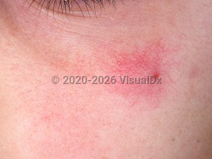
A spider angioma, also known as a spider vein or spider nevus, is the most prevalent of the telangiectases. Clinically, there is a central arteriole from which numerous small, twisted vessels radiate. The ascending central arteriole appears as a spider's body, and the radiating vessels resemble the spider's legs, hence the spider appearance that is visible on the skin.
This common benign acquired lesion usually appears spontaneously and is present in 40% of normal children up to age 8. Prevalence drops to about 10%-15% of healthy adults. Many women develop lesions during pregnancy or while taking oral contraceptives, likely due to high estrogen levels in their blood. These lesions usually disappear following parturition or cessation of the contraceptives.
Spider angiomas may be indicative of underlying systemic disease, especially when found in large numbers. Liver dysfunction due to hepatic cirrhosis or hepatic tumors impair the metabolism of estrogen, which may play a role in increased nevus formation. In addition, elevated levels of serum vascular endothelial growth factor (VEGF) and young age are predictive of spider angioma formation in patients with cirrhosis. Patients with alcoholic cirrhosis are more likely to develop spider angiomas than those with viral or idiopathic cirrhosis. In patients with diseases of the liver, regression of the nevus may occur following improvement of the underlying condition, although this is not usually so. They are also associated less frequently with thyrotoxicosis and in patients on estrogen therapy.
Spider angiomas usually appear on the upper half of the body, frequently on sun-exposed areas. It is very uncommon for lesions to occur below the level of the umbilicus. The lesion ranges in size from that of a pinhead to 2 cm.
A lobular capillary hemangioma (pyogenic granuloma) developing within a large spider angioma is a possible complication of these benign vascular malformations.
Spider angioma in Adult
Alerts and Notices
Important News & Links
Synopsis

Codes
ICD10CM:
I78.1 – Nevus, non-neoplastic
SNOMEDCT:
195382003 – Spider nevus
I78.1 – Nevus, non-neoplastic
SNOMEDCT:
195382003 – Spider nevus
Look For
Subscription Required
Diagnostic Pearls
Subscription Required
Differential Diagnosis & Pitfalls

To perform a comparison, select diagnoses from the classic differential
Subscription Required
Best Tests
Subscription Required
Management Pearls
Subscription Required
Therapy
Subscription Required
Drug Reaction Data
Subscription Required
References
Subscription Required
Last Reviewed:08/19/2023
Last Updated:08/20/2023
Last Updated:08/20/2023
 Patient Information for Spider angioma in Adult
Premium Feature
VisualDx Patient Handouts
Available in the Elite package
- Improve treatment compliance
- Reduce after-hours questions
- Increase patient engagement and satisfaction
- Written in clear, easy-to-understand language. No confusing jargon.
- Available in English and Spanish
- Print out or email directly to your patient
Upgrade Today

Spider angioma in Adult