Vestibular schwannomas, also known as acoustic neuromas, are intracranial tumors that arise from the Schwann cell sheath of the eighth cranial (vestibulocochlear) nerve. The incidence of vestibular schwannomas in the United States is 10-20 per 1 000 000 people. Vestibular schwannomas account for approximately 8% of intracranial tumors in adults and represent 80% of all tumors of the cerebellopontine angle. Ninety percent of tumors are unilateral, affecting the left and right sides equally. The median age at diagnosis is 50 years. The tumors are rare in children, except for patients with neurofibromatosis type 2. There is a 3:2 female to male ratio. All races and ethnicities are equally affected.
The most common presenting symptoms are one-sided hearing loss and/or tinnitus. Vertigo and disequilibrium are relatively uncommon presentations. As the tumor expands, it may compress the facial or trigeminal nerves, resulting in decreased facial sensation or facial muscle weakness. Late complications include cerebellar and brainstem dysfunction as a result of compression. Obstructive hydrocephalus is also possible due to effacement of the fourth ventricle.
On physical examination, the Weber and Rinne tests and formal audiogram may be used to uncover unilateral sensorineural hearing impairment. Neurologic examination may reveal defects of the fifth and seventh cranial nerves, such as a decreased or absent corneal reflex or hypoesthesia. Romberg and Hall-Pike tests are typically normal.
Patients with neurofibromatosis type 2 classically develop bilateral vestibular schwannomas at a young age. Other risk factors associated with the development of vestibular schwannomas include childhood exposure to low dose radiation and a history of parathyroid adenoma.
Vestibular schwannomas are typically slow-growing tumors. They usually grow at a rate of 2-4 mm/year but can occasionally grow as rapidly as 2 cm/year. If treated with current techniques, patients have a very good prognosis with minimal complications. However, if left untreated, these tumors eventually lead to lethal brain stem compression or intracranial hypertension.
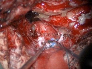
Vestibular schwannoma
Alerts and Notices
Important News & Links
Synopsis

Codes
ICD10CM:
D33.3 – Benign neoplasm of cranial nerves
SNOMEDCT:
126949007 – Acoustic neuroma
D33.3 – Benign neoplasm of cranial nerves
SNOMEDCT:
126949007 – Acoustic neuroma
Look For
Subscription Required
Diagnostic Pearls
Subscription Required
Differential Diagnosis & Pitfalls

To perform a comparison, select diagnoses from the classic differential
Subscription Required
Best Tests
Subscription Required
Management Pearls
Subscription Required
Therapy
Subscription Required
References
Subscription Required
Last Updated:04/08/2024