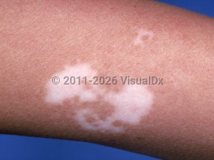
Vitiligo is a relatively common disorder characterized by acquired depigmentation of the skin resulting from the autoimmune destruction of melanocytes. Vitiligo manifests as patterned white skin patches arising anywhere on the body but more frequently in areas of recurrent trauma, particularly on the face, upper chest, hands, elbows, knees, axillae, and perineum.
The prevalence of vitiligo in childhood and adolescence is between 0.5 % and 2.16%. Vitiligo affects males and females equally. It has a bimodal age of onset, with about one-third of cases starting in childhood (mean age of around 10.3 years) and two-thirds of cases presenting in adulthood (mean age of 34 years). Congenital vitiligo is rare, but cases have been reported.
More than two-thirds of childhood vitiligo cases are classified as nonsegmental vitiligo, with a bilateral and symmetric distribution of lesions on the body. The disease course of nonsegmental vitiligo is more unpredictable, with progressive onset and multiple flares. The most prevalent types of nonsegmental vitiligo in children are vulgaris (42.49%), focal (27.21%), and acrofacial (17.8%). Segmental vitiligo typically involves a unilateral segment of the body. The segmental form disproportionately affects children as compared to adults and is less likely to be associated with coexisting autoimmune conditions.
While the precise etiology of vitiligo remains debated, the leading hypothesis implicates both genetic and environmental factors that lead to an innate immune response resulting in CD8+ T-cell activation, release of interferon-γ (IFN-γ), and an associated inflammatory cascade in keratinocytes via the JAK/STAT pathway.
Environmental factors include mechanical trauma and/or exposure to depigmenting agents.
While most vitiligo patients are otherwise healthy, an association with autoimmune thyroid dysfunction (hyperthyroidism or hypothyroidism) has been demonstrated. In new-onset vitiligo patients with systemic symptoms, thyroid screening with antithyroid peroxidase (TPO) antibody and a serum thyrotropin is recommended. Additional associations include endocrinopathies, such as diabetes mellitus and Addison disease, along with other autoimmune processes. Rarely, it may exist as part of polyglandular autoimmune syndrome, particularly a type III syndrome (eg, Hashimoto thyroiditis or alopecia areata and/or another organ-specific autoimmune disease). Vitiligo may accompany halo nevi. New-onset vitiligo may be seen in patients with metastatic melanoma. It can occur spontaneously and may herald metastatic disease, or it can be triggered by immunotherapy, such as with BRAF inhibitors or PD-1 inhibitors. In the latter setting, it is considered a good prognostic sign. Rarely, vitiligo may be associated with uveitis. An increased risk of developing sensorineural hearing loss has been identified in both adults and children with vitiligo. Additionally, there is an increased risk of mixed (sensorineural and conductive) hearing loss in children.
Vitiligo carries a psychosocial burden and negatively impacts the quality of life of affected children. Mental health conditions have been associated with vitiligo including anxiety, depression, panic disorder, and attention deficit hyperactivity disorder (ADHD).
Vitiligo in Child
See also in: External and Internal Eye,AnogenitalAlerts and Notices
Important News & Links
Synopsis

Codes
ICD10CM:
L80 – Vitiligo
SNOMEDCT:
56727007 – Vitiligo
L80 – Vitiligo
SNOMEDCT:
56727007 – Vitiligo
Look For
Subscription Required
Diagnostic Pearls
Subscription Required
Differential Diagnosis & Pitfalls

To perform a comparison, select diagnoses from the classic differential
Subscription Required
Best Tests
Subscription Required
Management Pearls
Subscription Required
Therapy
Subscription Required
Drug Reaction Data
Subscription Required
References
Subscription Required
Last Reviewed:09/17/2024
Last Updated:06/22/2025
Last Updated:06/22/2025
 Patient Information for Vitiligo in Child
Premium Feature
VisualDx Patient Handouts
Available in the Elite package
- Improve treatment compliance
- Reduce after-hours questions
- Increase patient engagement and satisfaction
- Written in clear, easy-to-understand language. No confusing jargon.
- Available in English and Spanish
- Print out or email directly to your patient
Upgrade Today

Vitiligo in Child
See also in: External and Internal Eye,Anogenital