Contents

SynopsisCodesLook ForDiagnostic PearlsDifferential Diagnosis & PitfallsBest TestsManagement PearlsTherapyDrug Reaction DataReferences

Information for Patients

View all Images (11)

Keratoacanthoma - Hair and Scalp
See also in: Overview,External and Internal Eye
Other Resources UpToDate PubMed

Keratoacanthoma - Hair and Scalp

See also in: Overview,External and Internal Eye
Contributors: Jennifer Lee MD, Jeffrey M. Cohen MD, Susan Burgin MD
Other Resources UpToDate PubMed

Synopsis

A keratoacanthoma is a rapidly growing, well-differentiated neoplasm of squamous epithelium. It is considered by many to be a low-grade squamous cell carcinoma (SCC). In contrast to classic SCCs, keratoacanthomas typically appear and grow rapidly over a few weeks and spontaneously involute and resolve within 6 months, leaving an atrophic scar. The immune system is thought to play a role in the spontaneous regression of keratoacanthomas.

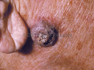
Keratoacanthomas are most commonly seen in individuals aged 60 years and older with lighter skin colors and a history of prolonged sun exposure. Men are more commonly affected than women.

Keratoacanthomas typically present as solitary, crater-shaped nodules measuring a couple centimeters in diameter, often with a central keratin plug on sun-exposed skin.

While solitary keratoacanthomas as described above are the most common presentation, there are variants. The giant type is usually larger and measures between 2-15 cm in diameter, the subungual type occurs underneath the nail, the mucosal type occurs on mucous membranes, and the keratoacanthoma centrifugum marginatum type has a more prominent horizontal growth pattern.

Risk factors for keratoacanthomas include ultraviolet radiation, human papillomavirus infection, immunosuppression, and certain medications. Patients on immunosuppressant medications tend to have more persistent and chronic keratoacanthomas. Patients taking medications such as BRAF inhibitors or hedgehog inhibitors have also been reported to develop keratoacanthomas. In addition, skin injury may be a predisposing factor, and there are reports of keratoacanthomas developing in sites of prior trauma, in surgical scars, after laser resurfacing, and following radiation therapy. In rare cases, keratoacanthomas may develop as part of a syndrome.
  • In Ferguson-Smith syndrome, patients develop multiple self-healing keratoacanthomas. It is often seen in younger individuals. It is a genetic condition inherited in an autosomal dominant manner.
  • In generalized eruptive keratoacanthomas of Grzybowski, patients develop hundreds to thousands of keratoacanthomas on both the skin and mucous membranes. These keratoacanthomas are frequently chronic and progressive.
  • In Muir-Torre syndrome, patients may develop keratoacanthomas along with sebaceous neoplasms and/or gastrointestinal or genitourinary malignancy.
  • In familial keratoacanthomas of Witten and Zak, patients develop multiple large and small keratoacanthomas; it is inherited in an autosomal dominant fashion.

Codes

ICD10CM:
L85.8 – Other specified epidermal thickening

SNOMEDCT:
254662007 – Keratoacanthoma

Look For

Subscription Required

Diagnostic Pearls

Subscription Required

Differential Diagnosis & Pitfalls

To perform a comparison, select diagnoses from the classic differential

Subscription Required

Best Tests

Subscription Required

Management Pearls

Subscription Required

Therapy

Subscription Required

Drug Reaction Data

Subscription Required

References

Subscription Required

Last Reviewed:03/06/2024
Last Updated:03/18/2024
Copyright © 2024 VisualDx®. All rights reserved.
Patient Information for Keratoacanthoma - Hair and Scalp
Print E-Mail Images (11)
Contributors: Medical staff writer
Premium Feature
VisualDx Patient Handouts
Available in the Elite package
  • Improve treatment compliance
  • Reduce after-hours questions
  • Increase patient engagement and satisfaction
  • Written in clear, easy-to-understand language. No confusing jargon.
  • Available in English and Spanish
  • Print out or email directly to your patient
Copyright © 2024 VisualDx®. All rights reserved.
Keratoacanthoma - Hair and Scalp
See also in: Overview,External and Internal Eye
A medical illustration showing key findings of Keratoacanthoma
Clinical image of Keratoacanthoma - imageId=213569. Click to open in gallery.  caption: 'A close-up of a violaceous nodule with a central keratotic core.'
A close-up of a violaceous nodule with a central keratotic core.
Copyright © 2024 VisualDx®. All rights reserved.